PART He-P 4040
REGISTRATION OF RADIATION MACHINES, MAGNETIC RESONANCE IMAGING MACHINES
AND PROVIDERS OF SERVICES
He-P 4040.01 Purpose. This part provides for:
(a) The evaluation of the need
for shielding for new and modified ionizing radiation facilities;
(b) The process for the
registration of ionizing radiation machines;
(c) The process for the
registration of magnetic resonance imaging (MRI) machines;
(d) The process for the
registration of persons providing radiation machine or MRI servicing, or services; and
(e) The process for the
approval of an out-of-state radiation or MRI machine to be brought into the
State of New Hampshire.
Source. #6827, eff 8-6-98; ss by #7919, eff
7-18-03; ss by #9945-A, eff 6-22-11; ss by #10864, eff 6-25-15
He-P 4040.02 Scope. This part shall apply to each person:
(a) In possession of an
ionizing radiation or MRI machine;
(b) Who wishes to dispose of or
make a change of ownership of a radiation or MRI machine;
(c) Who offers radiation or MRI
machine servicing or services; or
(d) Who wishes to bring a
radiation or MRI machine, from another state, into the State of New Hampshire.
Source. #6827, eff 8-6-98; ss by #7919, eff
7-18-03; ss by #9945-A, eff 6-22-11; ss by #10864, eff 6-25-15
He-P 4040.03 Shielding Plan
Review.
(a) Prior to new construction
or modification of existing installations utilizing ionizing radiation
machines, a review of floor plans, shielding specifications, and equipment
arrangements shall be completed as set forth in paragraphs (b)-(d) below to
ensure that, once operational, no individual will receive a dose in excess of
the limits prescribed in He-P 4020.
(b) All prospective and
existing registrants shall utilize the design services of a registered service
provider as described in He-P 4040.07.
(c) Each prospective or
existing registrant may request the department of health and human
services/radiological health section (DHHS/RHS) to provide an evaluation of an
existing shielding plan review or to offer technical advice on shielding
requirements for a radiation machine installation.
(d) The following information
shall be submitted to DHHS/RHS with any request for a shielding plan review:
(1) Distances where applicable,
such as:
a. From the source of radiation
to the patient;
b. From the source of radiation
to each point for which the shielding requirement is to be calculated; and
c. From the patient to each
point for which the shielding requirement is to be calculated;
(2) The normal location(s) of
the system's radiation port;
(3) The travel and traverse
limits of the radiation beam port(s);
(4) Direction(s) in which the
useful beam will be directed;
(5) Locations of any windows,
doors or other openings;
(6) The location of the
operator's booth;
(7) The location of the control
panel;
(8) The structural composition
and thickness or lead equivalent of all existing or planned walls, doors,
partitions, floor, and ceiling of the room(s) to be evaluated;
(9) The dimensions of the
room(s) to be evaluated;
(10) The type of occupancy of
all adjacent areas, including the space above and below the rooms(s), and, if
there is an exterior wall, the distance to the closest area(s) where it is
likely that an individual might be present;
(11) The make and model of the
equipment, the maximum technique factors as defined in He-P 4041, and the
output waveform;
(12) The type of examination(s)
or treatment(s) to be performed with the equipment; and
(13) Information on the
anticipated workload of the system(s) in milliampere seconds (mAs) per week, milliampere seconds being the product of the
x-ray tube current and the exposure time.
(e) After installation of a
radiation machine, the registrant shall keep the following records for review
during an inspection by DHHS/RHS until the registration is terminated:
(1) The maximum rated technique
factors, as defined in He-P 4041, of each machine;
(2) A scaled drawing of the
room in which each stationary radiation machine system is located indicating
the location of tubeheads, describing the use of each
area adjacent to the room, and specifying an estimation of the extent of
occupancy by an individual in such areas;
(3) The results of a survey for
radiation levels present at the operator's position and at pertinent points
outside the room at specified test conditions; and
(4) The shielding plan review
which shall include the type and thickness of materials, or lead equivalency,
of each protective barrier.
(f) After installation of an
MRI machine and until the registration is terminated, the registrant shall keep
the following records for review during an inspection by DHHS/RHS:
(1) A scaled drawing of the MRI
suite indicating the location of reception, patient, prescreening, control
room, MRI machine, fire extinguishers and equipment;
(2) An MRI suite safety
protocol which shall be reviewed:
a. Annually; and
b. After any change is made to
the MRI suite; and
(3) Documentation that the
local fire department is aware of the presence of MRI suite hazards in the
event of an emergency response.
Source. #6827, eff 8-6-98; ss by #7919, eff
7-18-03; ss by #9945-A, eff 6-22-11; ss by #10864, eff 6-25-15
He-P 4040.04 Registration of
Radiation or MRI Machines.
(a) Each person having
possession of any ionizing radiation or MRI machine, except those specifically
exempted in He-P 4040.06, or those applying for reciprocal recognition of an
out-of-state radiation or MRI machine in accordance with 4040.11, shall
register such machine with DHHS/RHS within 30 days after its acquisition.
(b) Each person leasing or
renting a radiation or MRI machine as defined in He-P 4041 shall be subject to
the machine registration requirements of He-P 4040.04.
(c) Application for
registration of a radiation or MRI machine shall be made on FORM DHHS/RHS A
“Application for Radiation or Magnetic Resonance Imaging Machine Registration”
(May, 2015) provided by DHHS/RHS.
(d) The registration period
shall begin on August 1 and end on July 31st of the following year.
(e) All registrations shall be
renewable as described in He-P 4040.09.
(f) No person having possession
of a radiation or MRI machine shall operate such machine or cause it to be
operated until it is registered with DHHS/RHS pursuant to He-P 4040.04.
(g) DHHS/RHS shall issue a
certificate of registration provided that the requirements of He-P 4040.04 and
the applicable fee payment required under He-P 4070.06 have been met.
(h) The registrant shall retain
the certificate of registration until it expires as per He-P 4040.08.
(i)
The registrant shall notify DHHS/RHS in writing within 15 days of any
change in the information submitted in the application for registration and
contained on the certificate of registration.
(j) The disposal or change of
ownership of any registered machine shall be made in accordance with He-P
4040.05.
(k)
Each registrant shall be responsible for radiation
protection in operations with each radiation machine.
(l) Each registrant shall
prohibit any person from furnishing radiation or MRI machine servicing or
services as described in He-P 4040.07 to a radiation or MRI machine or facility
until such person provides a current certificate of registration as evidence
that they are registered with DHHS/RHS as a provider of services.
(m) No person shall assemble,
install or use radiation or MRI machines unless such machines, when properly
placed in operation, meet the requirements of this chapter.
(n) Machines purchased during
the month of July shall be registered according to He-P 4040.04(a),
and shall be assessed the applicable fee set forth in He-P 4070 during
the month of August.
(o) In addition to the requirements set forth
in He-P 4040:
(1) All radiation and MRI
machine registrants shall follow the requirements set forth in He-P 4001
through He-P 4003, He-P 4019 through He-P 4022, He-P 4041 and He-P 4045;
(2) Registrants utilizing
analytical x-ray and radiation generating devices shall be subject to the
requirements of He-P 4043;
(3) Registrants utilizing
particle accelerators shall be subject to the requirements of He-P 4044;
(4) Registrants utilizing
radiation or MRI machines for all diagnostic purposes, including veterinarians
shall be subject to the requirements of He-P 4045 and He-P 4046;
(5) Registrants utilizing
therapeutic radiation machines shall be subject to the requirements of He-P
4047; and
(6) Registrants engaged in
industrial radiographic operations shall be subject to the requirements of He-P
4034.
Source. #6827, eff 8-6-98; ss by #7919, eff
7-18-03; amd by #9945-A, eff 6-22-11, (paras (a),
(c), & (d)-(j)); amd by #9945-B, eff 6-22-11, (para (b)); ss
by #10864, eff 6-25-15
He-P 4040.05 Disposal of or Change of Ownership of a
Radiation or MRI Machine.
(a) A machine
registration shall be non-transferable.
(b) A registrant shall submit a completed FORM
DHHS/RHS D “Certificate - Disposition or Change of Ownership of Radiation or
MRI Machine” (May, 2015) provided by DHHS/RHS when
modifying the possession of a registered machine as follows:
(1) Upon disposal of a registered machine either
by donation or disposal;
(2) Upon change of ownership of a business entity
with a registered machine(s);
(3) Upon replacement of a registered machine by a
registered service provider;
(4) Upon disposal of an originally registered
machine unable to be repaired or calibrated; or
(5) Upon the sale of a registered machine.
Source.
#10864, eff 6-25-15
He-P 4040.06 Exemptions.
(a) Any electronic equipment that produces
ionizing radiation incidental to its operation shall be exempt from the
requirements of this part provided that the dose equivalent rate averaged over
an area of 10 square centimeters does not exceed 5 μSv
(0.5 millirem) per hour at 5 centimeters from any accessible surface of such
equipment.
(b) Any radiation or MRI machine while in transit
or in storage incident thereto shall be exempt from the registration
requirements of this part.
(c) Domestic television receivers and video
display terminals shall be exempt from the requirements of this part.
(d) Permanently inoperable radiation or MRI
machines shall be exempt from the requirements of this part.
(e) Loaner machines, as defined in He-P 4041,
shall be exempt from the requirements of this part as long as
the loan does not exceed the 30 days. If
the loaner machine remains in the state after 30 days, the registrant shall
comply with He-P 4040.04, He-P 4040.05, and He-P 4070. The registrant shall submit written
notification to DHHS/RHS regardless of the duration of use of the loaner
machine. Notification shall include
registrant name, and the machine make, model, serial number and acquisition
date.
(f) Financial institutions that take possession
of a radiation or MRI machine as a result of foreclosure, bankruptcy, or other
default of payment shall be exempt from the requirements in this part to the
extent that they demonstrate that the unit is only operable for the sole
purpose of selling or leasing the unit.
(g) An individual employed by a registrant to
perform, on an in-house basis only, calibrations, equipment performance
evaluations, or minor repairs of that registrant’s diagnostic or industrial
radiation or MRI machines, shall be exempt from the service provider
registration requirements of this part, unless the individual also provides
these services to other registrants.
(h) The following shall not be exempt:
(1) Electron beam welders;
(2) Electron microscopes;
(3) Providers of radiation or MRI machines for
mobile services; and
(4) Providers of service for electronic equipment
in (a) above during testing or servicing.
(i) When the registrant of the portable radiation
machine, used solely for security-related, non-human imaging applications, is a
state agency or local government, the registrant shall be exempt from the
payment of the registration fee as required under He-P 4070.06. The registrant shall ensure that possession
and use of the radiation machine comply with all applicable requirements of
He-P 4000, New Hampshire Rules for the Control of Radiation.
Source. #6827, eff 8-6-98; ss by #7919, eff
7-18-03; ss by #9945-A, eff 6-22-11; ss by #10864, eff 6-25-15 (from He-P
4040.05); amd by#13280, eff 10-21-21
He-P 4040.07 Application for
Registration of Servicing and Services.
(a) Each person who is engaged
in the business of installing radiation or MRI machines, or
is engaged in the business of furnishing or offering to furnish radiation or
MRI machine servicing or services as described in He-P 4040.07(b) shall apply
for registration with DHHS/RHS prior to furnishing or offering to furnish such
services.
(b) Application for
registration of persons providing servicing or services shall be made on FORM DHHS/RHS SP “Application for
Registration as a Provider of Radiation or MRI Machine-Related Services” (May, 2015) provided
by DHHS/RHS.
(c) The applicant shall specify
each applicable type of service being offered, choosing from the following:
(1) Therapy unit services as
follows:
a. Radiation therapy physicist
as required by He-P 4047.04(k); or
b. Calibration and compliance
surveys of radiation therapy units;
(2) Installation and servicing
of radiation or MRI machines, therapeutic radiation machines or radiation
machines for other use and associated radiation or MRI machine components;
(3) Radiation protection
consultation or surveys;
(4) Design of shielding or
access controls for diagnostic, therapeutic or other facilities;
(5) Calibration of radiation or
MRI machines, measurement instruments, or radiation devices;
(6) Personnel dosimetry
services; or
(7) Distribution or sales of
radiation or MRI machines.
(d) Each person applying for
registration under He-P 4040.07 shall also provide the following:
(1) A description of the
minimum education, training, and experience that the applicant will require of each individual who will provide services under this
registration, as follows:
a. For installers, relative to
radiation safety and any applicable federal or state regulations pertaining to
the manufacture, assembly, installation, acceptance and quality control
testing, and functioning of radiation or MRI machines; and
b. For radiation protection
consultants and designers of shielding and access controls, relative to
radiation safety and protection, including radiation surveys and measurement,
instrumentation, shielding, and radiation protection engineering; and
c. For qualified experts, the
individual shall:
1. Be certified in the appropriate field by the
American Board of Radiology, the American Board of Health Physics, the American
Board of Medical Physics or the American Board of Nuclear Medicine Science; or
2. Hold a master’s or doctoral
degree in physics, biophysics, radiological physics, health physics, or medical
physics and have completed one year of documented, full time
training in the appropriate field and also one year of documented, full time work experience under the supervision of a
qualified expert in the appropriate field.
To meet this requirement, the individual shall have performed the tasks
required of a qualified expert during the year of work experience; or
3. Receive approval from
DHHS/RHS for specific activities if he or she has:
a. A bachelor’s degree in
physics, biophysics, radiological physics, health physics, or medical physics;
and
b Completed 6 years of
documented full time training and work experience in
the appropriate field;
(2) A complete list of all
individuals who will be providing services in New Hampshire under this
registration;
(3) A description of the
measuring instruments to be used for the type of service to be provided and the
frequency of calibration of those instruments; and
(4) An attestation, signed and
dated by the applicant, or management representative stating: “I certify that the applicant (1) understands and shall follow the relevant requirements
of the New Hampshire Rules for the Control of Radiation (He-P 4000)
applicable to the types of servicing or services to be provided under this
registration and (2) shall adhere and require adherence to them and good
radiation safety practices.”
(e) No registered service
provider shall perform services that are not specifically stated on the
certificate of registration issued by DHHS/RHS.
(f) DHHS/RHS shall issue a
certificate of registration provided that the requirements of He-P 4040.07 and
the fee required by He-P 4070.06 have been met.
(g) The registered service
provider shall retain the certificate of registration until it expires.
(h) Registration is renewable
as described in He-P 4040.09.
Source. #6827, eff 8-6-98; ss by #7919, eff
7-18-03; amd by #9945-A, eff 6-22-11, (paras (a)
& (c)-(e)); amd by #9945-B, eff 6-22-11, (para
(b)); ss by #10864, eff 6-25-15
He-P 4040.08 Expiration of Certificates of Registration.
(a) Each certificate of registration for
possession of a radiation or MRI machine shall expire at the end of the day on
the first July 31 after issuance of the certificate.
(b) Each certificate of registration for servicing and services shall expire at the end of the day on
the first July 31 after issuance of the certificate.
(c) The registration for any individual machine
shall terminate upon disposal, permanent removal or change of ownership, of the
machine as described in He-P 4040.05.
Source. #6827, eff 8-6-98; ss by #7919, eff
7-18-03; ss by #9945-A, eff 6-22-11; ss by #10864, eff 6-25-15 (from He-P
4040.07)
He-P 4040.09 Renewal of Certificates of Registration.
(a) Renewal of a certificate of registration for
possession of a radiation or MRI machine shall be made in writing prior to
August 1st of each year.
(b) The machine renewal application shall include
the following:
(1) Updated or changed information about the
radiation or MRI machine status since issuance of the most recent certificate
of registration, including any of the following:
a. Type of machine;
b. Location of machine;
c. Transfer of ownership
d. Transfer to alternate site;
e. Change in status from in use to storage;
f. Change in status from storage to in use; or
g. Addition of any radiation or MRI machine;
(2) A completed FORM DHHS/RHS A “Application for
Radiation or Magnetic Resonance Imaging Machine Registration” (May, 2015) as required in He-P 4040.04 if any changes have
been made;
(3) A completed FORM DHHS/RHS D
“Certificate-Disposition or Change of Ownership of Radiation or MRI Machine” (May, 2015) as required in He-P 4040.05, if applicable;
(4) A check made payable to “Treasurer- State of
New Hampshire” for the appropriate fee listed in He-P 4070.06; and
(5) An attestation signed and dated by the
registrant, or a certifying officer signing on behalf of the registrant,
attesting to the following: “I, the
registrant or certifying officer signing on behalf of the registrant,
understand and shall follow the relevant requirements of the New Hampshire
Rules for the Control of Radiation (He-P 4000) and confirm that all of the
information herein is complete and accurate.”
(c) Each prospective or renewing registrant who
fails to submit the registration fee within 30 days of the date that the
application and fee are due, shall be assessed an
administrative fine of $100 with the fee as described in He-P 4070.08.
(d) Renewal of registration as service providers
pursuant to He-P 4040.07 shall be made in writing prior to August 1st
of each year.
(e) The service provider application for a
renewal of registration shall include the following:
(1) A signed and dated renewal of a certificate
of registration for servicing and services shall be made on FORM DHHS/RHS-RSP
“Annual Renewal of Radiation or MRI Machine-Related Service Provider
Registration” (May, 2015) provided by DHHS/RHS;
(2) An attestation signed and dated by the
applicant, or management representative stating “I certify that the applicant
(1) understands and shall follow the relevant requirements of the New Hampshire
Rules for the Control of Radiation (He-P 4000) applicable to the types of
services or servicing to be provided under this registration and (2) shall
adhere and require adherence to them and good radiation safety practices.”; and
(3) A check made payable to “Treasurer- State of
New Hampshire” for the appropriate fee listed in He-P 4070.06.
Source. #6827, eff 8-6-98; ss by #7919, eff
7-18-03; ss by #9945-A, eff 6-22-11; ss by #10864, eff 6-25-15; ss by #10864,
eff 6-25-15
He-P 4040.10 Assembler and/or Transferor Obligation.
(a) Any person who sells, leases, transfers,
lends, disposes, assembles, or installs radiation or MRI machines shall
register as a service provider and notify DHHS/RHS within 15 days of:
(1) The name and address of persons
who have received these machines;
(2) The manufacturer, model, and serial number of
each radiation or MRI machine transferred; and
(3) The date of transfer of each machine, if
applicable.
(b) No person shall make, sell, lease, transfer,
lend, assemble, or install radiation or MRI machines or the supplies used in
connection with such machines unless such supplies and equipment, when properly
placed in operation, meet the requirements of this chapter.
Source. #6827, eff 8-6-98; ss by #7919, eff
7-18-03; ss by #9945-A, eff 6-22-11; ss by #10864, eff 6-25-15 (from He-P
4040.09)
He-P 4040.11 Reciprocal Recognition of Out-of-State
Radiation and MRI Machines.
(a) A person proposing to bring any radiation or
MRI machine into New Hampshire from another state, for any use, shall apply to
DHHS/RHS in writing at least 3 business days prior to use of the machine in the
state.
(b) Any radiation or MRI machine used for
demonstration purposes shall follow He-P 4040.11.
(c) Application for recognition of a person
proposing to bring an out-of-state machine into New Hampshire shall be made on
FORM DHHS/RHS-RP “Registration for Reciprocal Recognition of Out-of-State
Machines” (May, 2015) provided by DHHS/RHS.
(d) The applicant shall provide, with the
application, a fee equal to half the annual fee set out in He-P 4070.06, for
each radiation or MRI machine the applicant proposes to bring into the state
with the first application for that radiation or MRI machine in the calendar
year.
(e) The reciprocity fee required by (d) above
shall cover a period of one year from the time of application, at which time a
new fee submittal shall be required.
(f) In the event an applicant proposes
subsequently to bring into the state in the same
calendar year a machine with a higher annual fee, the applicant shall provide
with the application a fee equal to half the difference between the fees of the
2 types of machines.
(g) DHHS/RHS shall not grant approval to any
application for an out-of-state radiation or MRI machine requiring a fee, if
the fee does not accompany the application.
(h) DHHS/RHS shall refund the fee accompanying
any application requiring one if it does not grant approval for that
application.
(i) The applicant
shall apply in writing for a waiver to use an out-of-state radiation or MRI
machine with less than 3-business days prior written notification.
(j) DHHS/RHS shall grant a waiver of the
3-business day application limit on determining that adherence to the limit
would endanger an individual’s or the public’s health and safety.
(k) The person entering the state under
reciprocity shall:
(1) Comply with the rules in this chapter as they
relate to radiation or MRI machines, their operators, and operations of the
machines;
(2) Supply additional information as requested by
DHHS/RHS for the purposes of protecting public and worker health and safety and
ensuring the safe use of machines within the state; and
(3) Not operate an out-of-state machine within
the state under reciprocity in excess of 180 calendar
days per year.
(l) Any radiation or MRI machine shall be subject
to unannounced inspection by DHHS/RHS during the duration of the out-of-state
recognition of reciprocal registration.
Source. #6827, eff 8-6-98; ss by #7919, eff
7-18-03; ss by #9945-A, eff 6-22-11; ss by #10864, eff 6-25-15 (from He-P
4040.10)
He-P 4040.12 Radiation or MRI Machines in Storage.
(a) A radiation or MRI machine shall be
categorized as a machine in storage if it meets the following conditions:
(1) The machine is registered with DHHS/RHS in
accordance with the requirements of He-P 4040.04;
(2) The machine is physically disconnected from
any source of operating electric power in one or more of the following ways:
a. By the removal of its power cord or plug, if
so equipped;
b. By the removal of
its direct connection from the facility's electrical wiring system, if so
connected;
c. By the removal of its internal main fuse or
circuit breaker, if so equipped; or
d. By having its external circuit breaker or
electrical circuit disconnect locked in an electrically non-conducting
condition with a key-operated or combination lock; and
(3) The machine has at its operating control or
console a tag or label bearing the words "In storage.”
(b) The placement of a machine into the storage
status shall be recorded on FORM DHHS/RHS D “Certificate-Disposition or Change
of Ownership of Radiation or MRI Machine” (May, 2015)
which shall be submitted to DHHS/RHS.
Upon approval by DHHS/RHS, the status of the registered machine shall be
changed to “in storage” while it remains out of service.
(c) A registrant shall not put into service or
restore to service a machine registered as in storage until a completed FORM DHHS/RHS
D “Certificate-Disposition or Change of Ownership of Radiation or MRI Machine”
(May, 2015) has been submitted and the difference
between the fee for the machines in storage and the fee that would otherwise
apply for such machine, as set out in He-P 4070.06, has been paid.
(d) DHHS/RHS shall not require machines in
storage under the provisions of this section to meet the requirements in Parts
He-P 4041 through He-P 4047, for that machine type.
(e) A registered machine approved of as in
storage shall qualify for the fee specified in He-P 4070.06 for a machine in
storage.
Source. #6827, eff 8-6-98; ss by #7919, eff
7-18-03; ss by #9945-A, eff 6-22-11; ss by #10864, eff 6-25-15 (from He-P
4040.11)
He-P 4040.13 Waivers.
(a) DHHS/RHS has found that He-P 4040 applies to
a variety of conditions and uses, and that the strict application of these
rules may result in hardship or misapplication. The purpose of these waiver
procedures is to accommodate those situations where strict adherence would not
be in the best interest of the public.
(b) Any person who is or would be directly and
adversely affected by the strict application of a rule in He-P 4040 may request
a waiver, provided that such waiver does not contravene a federal statute or
regulation, state statutory requirement, or state or federal constitutional
requirement.
(c) Any person who wishes to request a waiver
shall submit a written request for a waiver.
(d) The request for a waiver shall include the
following information:
(1) The identification of the facility or
activity to which the request relates;
(2) The specific citation to the He-P 4040 rule
from which a waiver is sought;
(3) A full explanation of why a waiver is being
requested;
(4) A full explanation of any alternate
procedure, method, or other activity that is sought to be substituted for the
procedure, method, or other activity that is required by the rule from which a
waiver is being sought;
(5) The limit of duration requested
for the waiver; and
(6) A full explanation of why the person believes
that having the waiver granted will not adversely impact the public health or
safety, the environment, or property.
(e) All requests for a waiver shall be submitted
to DHHS/RHS.
(f) Where a request for a waiver relates to an
application for a registration, the person requesting the waiver shall submit
the request as part of such application, or as soon thereafter as the person
identifies the need for the request.
(g) DHHS/RHS shall grant a request for a waiver
if the requesting party demonstrates that:
(1) The waiver shall not result in an adverse
effect to public health or safety, the environment, or property; and
(2) Strict compliance with the applicable section
of He-P 4040 rule:
a. Will result in
an adverse effect to the public
health or safety, the environment, or property; or
b. Is technologically infeasible.
(h) DHHS/RHS shall send written notification to
the person requesting the waiver of the determination on the request. If the
waiver is submitted as part of the registration application and is granted, it
shall become part of the registration document.
Source. #10505, eff 1-11-14; ss by #10864, eff
6-25-15 (from He-P 4040.12)
PART
He-P 4041 USE OF RADIATION MACHINES:
DEFINITIONS
Source. #6827, eff 8-6-98; ss by #8692, INTERIM, eff
7-27-06, EXPIRES: 2-2-07; ss by #8808, eff 1-24-07; ss by #10806, eff 3-28-15;
ss by #14300, eff 7-1-25, EXPIRES: 7-1-35
He-P 4041.02 Definitions.
(a) “Accessible surface” means the external surface of
the enclosure or housing of the radiation-producing machine as provided by the
manufacturer.
(b) “Accessory component” means a component which is:
(1) Used with diagnostic x-ray systems, such as a
cradle or film changer, that is not necessary for the compliance of the system
with applicable provisions of He-P 4041 but which
requires an initial determination of compatibility with the system; or
(2) Necessary for the compliance of the system with
applicable provisions of He-P 4041, but which may be interchanged with similar
compatible components without affecting the system’s compliance, such as one of
a set of interchangeable beam-limiting devices; or
(3) Compatible with all x-ray systems with which it may
be used and that does not require compatibility or installation instructions,
such as a tabletop cassette holder.
(c) “Added filtration” means any filtration which is in
addition to the inherent filtration.
(d) “Air kerma (K)”
means the kinetic energy released in air by ionizing radiation,
and is determined as the quotient of dE by dM, where dE is the sum
of the initial kinetic energies of all the charged particles liberated by
uncharged particles in air of mass dM, and is
expressed in the SI unit of joule per kilogram, and the special name for the
unit of kerma is “gray” (Gy).
(e) “Air kerma rate
(AKR)” means the air kerma per unit time.
(f) “Aluminum equivalent” means the thickness of type
1100 aluminum alloy for which the nominal chemical composition is 99.00 percent
minimum aluminum, 0.12 percent copper which affords the same attenuation, under
specified conditions, as the material in question.
(g) “Annual” means a period of
time or an event happening once a year.
(h) “Articulated joint” means a joint between 2
separate sections of a tabletop which provides the capacity of one of the
sections to pivot on the line segment along which the sections join.
(i) “Assembler” means any
person engaged in the business of assembling, replacing, or installing one or
more components into an x-ray system or subsystem and includes the owner of an
x-ray system or his or her employee or agent who assembles components into an
x-ray system that is subsequently used to provide professional or commercial
services.
(j) “Attenuation” means the reduction of exposure rate
upon passage of radiation through matter.
(k) “Attenuation block” means a block or stack of type
1100 aluminum alloy, or aluminum alloy having equivalent attenuation, with
dimensions 20 centimeters (cm) or larger by 20 cm or larger by 3.8 cm, that is
large enough to intercept the entire x-ray beam.
(l) “Automatic exposure control (AEC)” means a device
which automatically controls one or more technique factors in order to obtain at a pre-selected location(s) a
required quantity of radiation. The term includes devices such
as phototimers and ion chambers.
(m) “Automatic exposure rate control (AERC)” means a
device which automatically controls one or more technique factors in order to obtain at a pre-selected location(s) a
required quantity of radiation.
(n) “Backscatter” means the scattering of radiation or
particles in a direction opposite to that of the incident radiation due to
reflection from particles of the medium traversed.
(o) “Backscatter system” means a security screening
system that makes use of radiation scattered or deflected from an object or
person to form an image of the scattering object or person.
(p) “Beam axis” means a line from the source through
the center of the x-ray field.
(q) “Beam-limiting device” means a device which
provides a means to restrict the dimensions of the x-ray field.
(r) “Beam monitoring system” means a system designed to
detect and measure the radiation present in the useful beam.
(s) “Bone densitometry system” means a medical device
which uses electronically-produced ionizing radiation
to determine the density of bone structures of human patients.
(t) “Calibration” means:
(1) The determination of the response or reading of an
instrument relative to a series of known radiation values over the range of the
instrument;
(2) The determination of the radiation dose or exposure
rate at a designated distance from a radiation source under specified
conditions of measurement;
(3) To check, adjust, or systematically standardize to
graduations of a quantitative measuring instrument; and
(4) To check, adjust or systematically bring
radiation-producing equipment into manufacturer’s specifications.
(u) “C-arm fluoroscope” means a fluoroscopic x-ray
system in which the image receptor and x-ray tube housing assembly are
connected or coordinated to maintain a spatial relationship. A C-arm
fluoroscope system allows a change in the direction of the beam axis with
respect to the patient without moving the patient.
(v) “Cantilevered tabletop” means a tabletop designed
such that the unsupported portion can be extended at least 100 centimeters (cm)
beyond the support.
(w) “Cassette holder” means a device, other than a spot
film device, that supports and/or fixes the position of an image receptor
during a radiographic exposure.
(x) “Cephalometric device” means a device intended for
the radiographic visualization and measurement of the dimensions of the human
head.
(y) “Certified components” mean components of x-ray
systems which are subject to regulations promulgated under Public Law 90-602,
the Radiation Control for Health and Safety Act of 1968.
(z) “Certified system” means any x-ray system which has
one or more certified component(s).
(aa) “Changeable filters” means any filter, exclusive
of inherent filtration, which can be removed from the useful beam through any
electronic, mechanical, or physical process.
(ab) “Coefficient of variation” or “C” means the ratio
of the standard deviation to the mean value of a population of observations. It
is estimated using the following equation:

where:
s = Standard deviation of the population;
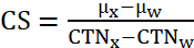 =
Mean value of observations in sample;
xi = The ith observation
sampled; and
n = Number of observations sampled.
(ac) “Computed radiography (CR)” means a projection
x-ray imaging method in which a cassette houses a phosphor plate rather than
photographic film. This photostimulable phosphor-coated plate
captures a latent image when exposed to x-rays and, when processed, releases
light that is converted to a digital image.
(ad) “Computed tomography (CT)” means the production of
a tomogram by the acquisition and computer processing of x-ray transmission
data.
(ae) “Computed tomography dose index (CTDI)” means the
integral from -7T to +7T of the dose profile along a line perpendicular to the
tomographic plane divided by the product of the nominal tomographic section
thickness and the number of tomograms produced in a single scan, that is:
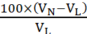
where:
z = Position along a line perpendicular to the tomographic plane;
D(z) = Dose at position z;
T = Nominal tomographic section thickness; and
n = Number of tomograms produced in a single scan.
This
definition assumes that the dose profile is centered around z = 0 and that, for
a multiple tomogram system, the scan increment between adjacent scans is nT.
(af) “Collimator” means a
device, as referenced in He-P 4043-4047, for restricting the useful radiation
in one or more directions.
(ag) “Cone beam computed tomography (CBCT)” means a
digital volume tomography method used in some imaging applications using 2 dimensional digital detector arrays, and a cone-shaped
x-ray beam, instead of a fan-shaped beam, that rotates around the patient to
generate a high resolution, 3D image, with high geometric
accuracy. Reconstruction algorithms can be used to generate images
of any desired plane.
(ah) “Contact therapy system” means an x-ray system
used for therapy with the x-ray tube port placed in contact with or within 5
centimeters of the surface being treated.
(ai) “Contrast resolution” means the ability to detect,
image, and display similar tissues or materials such as gray-white matter and
liver-spleen.
(aj) “Contrast scale (CS)”
means the change in the linear attenuation coefficient per CTN relative to
water, that is:

where:
μx =
Linear attenuation coefficient of the material of interest;
μw =
Linear attenuation coefficient of water;
CTNx= CTN
of the material of interest; and
CTNw= CTN
of water.
(ak) “Control panel” means
that part of the x-ray control upon which are mounted the switches, knobs,
pushbuttons, and other hardware necessary for manually setting the technique
factors.
(al) “Cooling curve” means the graphical relationship
between heat units stored and cooling time.
(am) “CT conditions of operation” means all selectable
parameters governing the operation of a CT x-ray system including, but not
limited to, nominal tomographic section thickness, filtration, and the
technique factors as defined in He-P 4041.02(fd).
(an) “CT dosimetry phantom” means the phantom used for
determination of the dose delivered by a CT x-ray system.
(ao) “CT gantry” means the
tube housing assemblies, beam-limiting devices, detectors, and the supporting
structures, frames, and covers which hold and/or enclose these components.
(ap) “CT number (CTN)” means the number used to
represent the x-ray attenuation associated with each elemental area of the CT
image.

where:
k = A constant, a normal value of 1,000
when the Hounsfield scale of CTN is used;
μx =
Linear attenuation coefficient of the material of interest; and
μw =
Linear attenuation coefficient of water.
(aq) “CT scan” means the
complete process of collecting x-ray transmission data for
the production of a tomogram. This includes data
collected simultaneously during a single scan for the
production of one or more tomograms.
(ar) “CT scan increment”
means the amount of relative displacement of the patient with respect to the CT
system between successive scans measured along the direction of the
displacement.
(as) “CT scan time” means the time between the
beginning and end of the x-ray transmission data accumulation for a CT scan.
(at) “Cumulative air kerma”
means the total air kerma accrued from the
beginning of an examination or procedure and includes all contributions from
fluoroscopic and radiographic irradiation.
(au) “Dead-man switch” means a switch so constructed
that a circuit closing contact can be maintained only by continuous pressure on
the switch by the operator.
(av) “Demonstration” means a showing of the merits of a
machine to a prospective customer.
(aw) “Diagnostic imaging system” means an assemblage of
components for the generation, emission, reception,
transformation, storage, and visual display of the resultant image.
(ax) “Diagnostic source assembly” means the tube
housing assembly with a beam-limiting device attached.
(ay) “Diagnostic x-ray system” means an x-ray system
designed for irradiation of any part of the human or animal body for the
purpose of diagnosis or visualization.
(az) “Diagnostic x-ray
imaging system” means an assemblage of components for the generation, emission
and reception of x-rays and the transformation, storage and visual
display of the resultant x-ray image.
(ba) “Digital radiography
(DR)” means an x-ray imaging method or radiography which produces a digital
rather than film projection image. The term includes both CR and
DDR.
(bb) “Direct digital radiography (DDR”) means an x-ray
imaging method in which a digital sensor, rather than photographic film or
phosphor plate, is used to capture an x-ray image. DDR is a
cassette-less imaging method providing faster acquisition time than
cassette-based CR, using an electronic sensor that converts x-rays to
electronic signals, current or charge, when exposed to x-rays.
(bc) “Direct scattered
radiation” means that scattered radiation which has been deviated in direction
only by materials irradiated by the useful beam.
(bd) “Dose area product (DAP)” means the product of the
air kerma and the area of the irradiated
field and is measured in Gy-cm2, so it does not change with the
distance from the x-ray tube. The term includes the term kerma-area product (KAP).
(be) “Dose profile” means the dose as a function of
position along a line.
(bf) “Elemental area” means
the smallest area within a tomogram for which the x-ray attenuation properties
of a body are depicted.
(bg) “Elemental area” means the smallest area
within a tomogram for which the x-ray attenuation properties of a body are
depicted.
(bh) “Emergency procedure”
means the written pre-planned steps to be taken in the event of actual or
suspected exposure of an individual in excess of
administrative or regulatory limits. This procedure includes the names and
telephone numbers of individuals to be contacted as well as directives for
processing the film badge or other personnel monitoring devices.
(bi) “Entrance skin exposure (ESE)” means x-ray
exposure of the skin, expressed in mR or
air kerma (Gy).
(bj) “Equipment” means “x-ray
equipment” as defined in He-P 4041.02(ft).
(bk) “Exposure rate” means the amount of ionization
produced or incident per unit of time.
(bl) “Field emission
equipment” means equipment which uses an x-ray tube in which electron emission
from the cathode is due solely to the action of an electric field.
(bm) “Filter” means material placed in the useful beam
to absorb preferentially selected radiations.
(bn) “Fluoroscopic
air kerma display devices” means separate
devices, subsystems or components that provide the display of AKR and
cumulative air kerma. They include
radiation detectors, if any, electronic and computer components, associated
software and data displays.
(bo) “Fluoroscopic imaging assembly” means the
image receptor(s), the image intensifier, the spot-film device, electrical
interlocks, and structural material providing linkage between the image
receptor and diagnostic source assembly as a subsystem in which x-ray photons
produce a fluoroscopic image.
(bp) “Fluoroscopic
irradiation time” means the cumulative duration during an examination or
procedure of operator-applied continuous pressure to the device, enabling x-ray
tube activation in any fluoroscopic mode of operation.
(bq) “Fluoroscopic procedure”
means the production and display of serial x-ray images for the purpose of
observing real-time motion of anatomical structures.
(br) “Fluoroscopy” means a technique for generating
x-ray images and presenting them simultaneously and continuously as visible
images.
(bs) “Focal spot” means the actual focal spot area
projected on the anode of the x-ray tube bombarded by the electrons accelerated
from the cathode and from which the useful beam originates.
(bt) “General purpose
radiographic x‑ray system” means any radiographic x-ray system which, by
design, is not limited to radiographic examination of specific anatomical
regions.
(bu) “Gonad shield” means a
protective barrier for the testes or ovaries.
(bv) “Half-value layer (HVL)” means the thickness of
specified material which attenuates the beam of radiation to an extent such
that the exposure rate is reduced to one-half of its original value and for
which the contribution of all scattered radiation, other than any which might
be present initially in the beam concerned, is deemed to be excluded.
(bw) “Hand-held x-ray
equipment” means x-ray equipment that is designed to be hand-held during
operation.
(bx) “Healing arts
screening” means the testing of human beings using x-ray machines for the
detection or evaluation of health indications when such tests are not
specifically and individually ordered by a licensed practitioner of the healing
arts legally authorized to prescribe such x-ray tests for the purpose of
diagnosis or treatment.
(by) “Heat unit” means a unit of energy equal to the
product of the peak kilovoltage, milliamperes, and seconds.
(bz) “Image intensifier”
means a device, installed in its housing, which instantaneously converts an
x-ray pattern into a corresponding light image of higher intensity.
(ca) “Image quality” means the exactness of a
2-dimensional representation of the patient’s anatomy on the radiographic
image.
(cb) “Image receptor” means
any device, such as a fluorescent screen or radiographic film, which transforms
incident x-ray photons either into a visible image or into another form which
can be made into a visible image by further transformations.
(cc) “Image receptor support device” means, for
mammographic systems, that part of the system designed to support the image
receptor in a horizontal plane during mammography.
(cd) “Inherent filtration”
means the filtration of the useful beam provided by the permanently installed
components of the tube housing assembly.
(ce) “Interventional procedures” means procedures
that utilize imaging for guidance. Imaging includes, but is not
limited to, fluoroscopy and CT scan.
(cf) “Irradiation” means the
exposure of matter to ionizing radiation.
(cg) “Isocenter” means the center of the smallest
sphere through which the beam axis passes when the equipment moves through a
full range of rotations about a common center.
(ch) “Kerma” means the quantity as defined by the
International Commission on Radiation Units and
Measurements. The kerma, K, is the
quotient of dEtr by dm,
where dEtr is the sum of the
initial kinetic energies of the all charged
particles liberated by the uncharged particles in mass dm of material thus K=dEtr/dm in units of J/kg, where the special name
for the unit of kerma is gray
(Gy). When the material is air, the quantity is referred to as
“air kerma.”
(ci) “Kilovolts peak (kVp)”
means “peak tube potential”.
(cj) “kV” means kilovolts,
which is 1,000 volts.
(ck) “kWs” means
kilowatt-second which is equivalent to 103 watt-second where 1
watt-second = kV × mA × second.
(cl) “Last image hold (LIH) radiograph” means an image
obtained either by retaining one or more fluoroscopic images, which can be
temporarily integrated, at the end of a fluoroscopic exposure or by initiating
a separate and distinct radiographic exposure automatically and immediately in
conjunction with termination of the fluoroscopic exposure.
(cm) “Lateral fluoroscope”
means the x-ray tube and image receptor combination in a biplane system
dedicated to the lateral projection. It consists of the lateral
x-ray tube housing assembly and the lateral image receptor that are fixed in
position relative to the table with the x-ray beam axis parallel to the plane
of the table.
(cn) “Lead equivalent” means
the thickness of lead affording the same attenuation, under specified
conditions, as the material in question.
(co) “Leakage radiation”
means radiation emanating from the diagnostic or therapeutic source assembly
other than the useful beam and radiation produced when the exposure switch or
timer is not activated.
(cp) “Leakage technique
factors” means the technique factors associated with the diagnostic or
therapeutic x-ray source assembly which are used in measuring leakage and are
defined as follows:
(1) For diagnostic source assemblies intended for
capacitor energy storage equipment, the maximum-rated peak tube potential and
the maximum-rated number of exposures in a hour
for operation at the maximum-rated peak tube potential with the quantity of
charge per exposure being 10 millicoulombs or the minimum obtainable from the
unit, whichever is larger;
(2) For diagnostic source assemblies intended for field
emission equipment rated for pulsed operation, the maximum-rated peak
tube potential and the maximum-rated number of x-ray pulses in an
hour for operation at the maximum-rated peak tube potential; and
(3) For all other diagnostic source assemblies, the
maximum-rated peak tube potential and the maximum-rated continuous
tube current for the maximum-rated peak tube potential.
(cq) “Lease” means a machine
that is possessed and used for a specified term in exchange for
consideration. The term includes “rental.”
(cr) “Light field” means that
area of the intersection of the light beam from the beam-limiting device and
one of the set of planes parallel to and including the plane of the image
receptor, whose perimeter is the locus of points at which the illumination is
one-fourth of the maximum in the intersection.
(cs) “Line-voltage regulation” means the difference
between the no-load and the load line potentials expressed as a percent of the
load line potential calculated using the following equation:
Percent
line-voltage regulation = 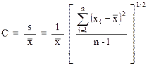
where:
VN = No-load line potential; and
VL = Load line potential.
(ct) “Loaner” means a machine
that is a replacement for a registered machine that is being repaired,
calibrated, or both. A loaner shall not include a machine that is
purchased as a replacement for a registered machine.
(cu) “mA” means milliampere, which is 1/1,000 of an
ampere.
(cv) “Magnetic resonance imaging scanner” means a
diagnostic tool using a combination of magnetic fields and radio frequency
waves to create cross-sectional images of the body.
(cw) “Magnetic resonance
imaging (MRI)” means a non-invasive diagnostic procedure used to create
cross-sectional images of the body by use of magnetic fields and radio
frequency fields.
(cx) “Mammo-tomo” means
breast tomosynthesis in which the x-ray tube moves in an arch during the
exposure. The term includes “three dimensional breast
imaging.”
(cy) “mAs” means milliampere
second, which is 1/1,000 of an ampere second.
(cz) “Maximum line current” means the
root-mean-square current in the supply line of an x-ray machine operating at
its maximum rating.
(da) “Micro computed tomography” (Micro CT)
means a scanning method that uses x-rays to create cross-sections of a
physical object that can be used to recreate a virtual, 3-dimensional
model. The pixel sizes of the cross-sections generated with this
method are in the micrometer range. The term includes “high-resolution
x-ray tomography.”
(db) “Mobile MRI unit” means
a magnetic resonance imaging scanner that is approved by the federal Food and
Drug Administration for the provision of diagnostic MRI services and which is
physically located in a vehicle such as a self-contained van or tractor trailer
for the purpose of transporting the magnetic resonance imaging scanner from one
site to another.
(dc) “Mobile x-ray equipment” means x-ray equipment mounted on a
permanent base with wheels and/or casters for moving while completely
assembled.
(dd) “Multiple tomogram system” means a computed
tomography x-ray system which obtains x-ray transmission data simultaneously
during a single scan to produce more than one tomogram.
(de) “Noise” means the standard deviation of the
fluctuations in CTN expressed as a percentage of the attenuation coefficient of
water and is calculated using the following equation:

where:
CS= Contrast scale;
μw =
Linear attenuation coefficient of water; and
s = Standard deviation of the CTN of picture elements in a
specified area of the CT image.
(df) “Nominal tomographic
section thickness” means the full width at half-maximum of the sensitivity
profile taken at the center of the cross-sectional volume over which x-ray
transmission data are collected.
(dg) “O-Arm” means a mobile fluoroscope, operated as a
C-arm fluoroscope, which is opened and positioned over the area of interest on
the patient being examined then closed around the patient to facilitate
360-degree x-ray imaging.
(dh) “Patient” means an individual or animal subjected
to healing arts examination, diagnosis, or treatment.
(di) “Peak tube potential” means the maximum value of
the potential difference across the x-ray tube during an exposure.
(dj) “Phantom” means a volume
of material with an atomic number (Z) and density similar
to biological tissue, which behaves in a manner similar
to tissue with respect to the attenuation and scattering of radiation.
(dk) “Physician assistant” means a person licensed as a
physician assistant by the New Hampshire board of medicine.
(dl) “Phototimer” means a method for controlling
radiation exposure to image receptors by the amount of radiation which reaches
a radiation monitoring device(s) that is part of an electronic circuit which
controls the duration of time the tube is activated.
(dm) “Picture element” means an elemental area of a
tomogram.
(dn) “Portable x-ray equipment” means x-ray equipment
designed to be hand carried.
(do) “Position indicating device (PID)” means a device
on dental x-ray equipment used to indicate the beam position and to establish a
definite source-surface (skin) distance. It may or may not
incorporate or serve as a beam-limiting device.
(dp) “Positive beam
limitation (PBL)” means the automatic or semi-automatic adjustment of an x-ray
beam to the size of the selected image receptor, whereby exposures cannot be
made without such adjustment.
(dq) “Primary protective
barrier” means the material, excluding filters, placed in the useful beam, for
protection purposes, to reduce the radiation exposure.
(dr) “Protective apron” means an apron made of
radiation absorbing materials used to reduce radiation exposure.
(ds) “Protective barrier” means a barrier of radiation
absorbing material(s) used to reduce radiation exposure.
(dt) “Protective glove” means a glove of radiation
absorbing materials used to reduce radiation exposure.
(du) “Pulsed mode” means operation of the x-ray system
such that the x-ray tube current is pulsed by the x-ray control to produce one
or more exposure intervals of duration less than one-half second.
(dv) “Quality assurance” means a program providing for
verification by written procedures such as testing, auditing, and inspection to
ensure that deficiencies, deviations, defective equipment, or unsafe practices,
or a combination thereof, relating to the use,
disposal, management, or manufacture of radiation devices are
identified, promptly corrected and reported to the appropriate regulatory
authorities.
(dw) “Qualified expert” means
an individual who has demonstrated to the satisfaction of DHHS/RHS that such
individual possesses the knowledge, skills, and training to measure
ionizing radiation to evaluate radiation parameters, to evaluate safety
techniques and to advise regarding radiation protection needs as described in
He-P 4040.
(dx) “Radiation detector” means a device which in the
presence of radiation provides a signal or other indication suitable for use in
measuring one or more quantities of incident radiation.
(dy) “Radiation therapy
simulation system” means a radiographic or fluoroscopic x-ray system intended
for localizing the volume to be exposed during radiation therapy and confirming
the position and size of the therapeutic irradiation field.
(dz) “Radiograph” means an
image receptor on which the image is created directly or indirectly by an x-ray
pattern and results in a permanent record.
(ea) “Radiographic imaging
system” means any system whereby a permanent or semi-permanent image is
recorded on an image receptor by the action of ionizing radiation.
(eb) “Rated line voltage”
means the range of potentials, in volts, of the supply line specified by the
manufacturer at which the x-ray machine is designed to operate.
(ec) “Rated output current”
means the maximum allowable load current of the x-ray high-voltage generator.
(ed) “Rating” means the operating limits as specified
by the component manufacturer.
(ee) “Recording” means producing a permanent form of an
image resulting from x-ray photons.
(ef) “Reference plane” means
a plane which is displaced from and parallel to the tomographic plane.
(eg) “Resolution” means the
ability to image 2 separate objects and visually distinguish one from the
other.
(eh) “Response time” means the time required for an
instrument system to reach 90 percent of its final reading when the radiation‑sensitive
volume of the instrument system is exposed to a step change in radiation flux
from zero sufficient to provide a steady state mid-scale reading.
(ei) “Scan” means the
complete process of collecting x-ray transmission data for
the production of a tomogram which may be collected simultaneously
during a single scan for the production of one or more
tomograms.
(ej) “Scan increment” means
the amount of relative displacement of the patient with respect to the CT x-ray
system between successive scans measured along the direction of such
displacement.
(ek) “Scan sequence” means a pre-selected set of two or
more scans performed consecutively under pre-selected CT conditions of
operation.
(el) “Scan time” means the period
of time between the beginning and end of x-ray transmission data
accumulation for a single scan.
(em) “Scattered radiation”
means radiation that, during passage through matter, has been deviated in
direction.
(en) “Secondary dose
monitoring system” means a system that will terminate irradiation in the event
of a failure of the primary system.
(eo) “Secondary protective
barrier” means a barrier sufficient to attenuate the stray radiation to the
required degree.
(ep) “Shutter” means a device attached to the tube
housing assembly which can intercept the entire cross-sectional area of the
useful beam and which has a lead equivalency not less
than that of the tube housing assembly.
(eq) “Single tomogram system” means a CT x-ray system
which obtains x-ray transmission data during a scan to produce a single
tomogram.
(er) “Spatial frequency” means a measure of the changes
in tissue attenuation characteristics. Abrupt changes have high spatial
frequency, such as bone-lung interface, and gradual changes, such as
liver-spleen interface, have low spatial frequency. Spatial frequency is
expressed as line pair per millimeter (lp/mm).
(es) “Spatial resolution” means the ability to image
anatomical structures or small objects that have high subject contrast, such as
bone versus soft tissue.
(et) “Solid state x-ray imaging device” means an
assembly, typically in a rectangular panel configuration, that intercepts x-ray
photons and converts the photon energy into a modulated electronic signature
representative of the x-ray intensity over the area of the imaging
device. The electronic signal is then used to create an image for
display, storage or both.
(eu) “Source” means the focal
spot of the x-ray tube.
(ev) “Source-image receptor
distance (SID)” means the distance from the source to the center of the input
surface of the image receptor.
(ew) “Source-skin distance
(SSD)” means the distance from the source to the center of the entrant x-ray
field in the plane tangent to the patient skin surface.
(ex) “Spot check” means an abbreviated calibration
procedure which is performed to assure that a previous calibration continues to
be valid.
(ey) “Spot film” means a
radiograph which is made during a fluoroscopic examination to permanently
record conditions which exist during that fluoroscopic procedure.
(ez) “Spot-film device” means
a device intended to transport and/or position a radiographic image receptor
between the x-ray source and fluoroscopic image receptor and includes a device
intended to hold a cassette over the input end of an image intensifier for the
purpose of making a radiograph.
(fa) “Stationary x-ray equipment” means x-ray equipment
which is installed in a fixed location.
(fb) “Stray radiation” means the sum of leakage and
scattered radiation.
(fc) “Supervising physician” means a physician who
holds a current license issued by the New Hampshire board of medicine and who
supervises all professional activities of a physician assistant.
(fd) “Technique factors”
means the following conditions of operation:
(1) For capacitor energy storage equipment, peak tube
potential in kV and quantity of charge in mAs;
(2) For field emission equipment rated for pulsed
operation, peak tube potential in kV, and number of x-ray pulses;
(3) For CT x-ray systems designed for pulsed operation,
peak tube potential in kV, scan time in seconds, and either tube current in mA,
x-ray pulse width in seconds, and the number of x-ray pulses per scan, or the
product of tube current, x-ray pulse width, and the number of x-ray pulses
in mAs;
(4) For CT x-ray systems not designed for pulsed
operation, peak tube potential in kV, and either tube current in mA and scan
time in seconds, or the product of tube current and exposure time in mAs and the scan time when the scan time and exposure
time are equivalent; and
(5) For all other equipment, peak tube potential in kV,
and either tube current in mA and exposure time in seconds, or the product of
tube current and exposure time in mAs.
(fe) “Tenth-value layer
(TVL)” means the thickness of a specified material which attenuates x-radiation
or gamma radiation to an extent such that the air kerma rate,
exposure rate, or absorbed dose rate is reduced to one-tenth of the value
measured without the material at the same point.
(ff) “Termination of irradiation” means the stopping of
irradiation in a fashion which will not permit continuance of irradiation
without the resetting of operating conditions at the control panel.
(fg) “Transmission system”
means a security screening system using the conventional means of radiographic
imaging in which x-rays pass through a target, such as a person or object and
create shadow-grams of enclosed objects, such as contraband, based on their
radiation attenuating properties.
(fh) “Tomogram” means the
depiction of the x-ray attenuation properties of a section through the body.
(fi) “Tomographic plane” means that geometric plane
which is identified as corresponding to the output tomogram.
(fj) “Tomographic section” means the volume of an
object whose x-ray attenuation properties are imaged.
(fk) “Tube” means an x‑ray
tube, unless otherwise specified.
(fl) “Tube housing assembly”
means the tube housing with tube installed to include the high-voltage and/or
filament transformers and other appropriate elements when such are contained
within the tube housing.
(fm) “Tube rating chart”
means the set of curves which specify the rated limits of operation of the tube
in terms of the technique factors.
(fn) “Useful beam” means the
radiation which passes through the tube housing port and the aperture of the
beam-limiting device when the exposure switch or timer is activated.
(fo) “Variable-aperture
beam-limiting device” means a beam-limiting device which has capacity for
stepless adjustment of the x-ray field size at a given SID.
(fp) “Visible area” means
that portion of the input surface of the image receptor over which incident
x-ray photons are producing a visible image.
(fq) “Wedge filter” means an
added filter effecting continuous progressive attenuation on all or part of the
useful beam.
(fr) “X-ray diffraction
(XRD)” means the analysis of the characteristic atomic pattern of x-rays
scattered when the primary beam from the machine strikes the sample.
(fs) “X-ray exposure control” means a device,
switch, button or other similar means by which an operator initiates
and/or terminates the radiation exposure and may include such associated
equipment as timers and back-up timers.
(ft) “X-ray equipment” means an x-ray system,
subsystem, or component thereof.
(fu) “X-ray field” means that area
of the intersection of the useful beam and any one of the set of planes
parallel to and including the plane of the image receptor, whose perimeter is
the locus of points at which the exposure rate is one-fourth of the maximum in
the intersection.
(fv) “X-ray
fluorescence (XRF)” means the analysis of the characteristic x-rays created
when the primary beam from the machine strikes the sample.
(fw) “X-ray
high-voltage generator” means a device which transforms electrical energy from
the potential supplied by the x-ray control to the tube operating potential and
may include a means for transforming alternating current to direct current,
filament transformers for the x-ray tube(s), high-voltage switches, electrical
protective devices, and other appropriate elements.
(fx) “X-ray
reciprocity” means any business entity proposing to bring a radiation machine
into this state from another state for operation only by the business entity,
except in areas of exclusive federal jurisdiction, for a period not in excess of 180 days in any calendar year.
(fy) “X-ray
subsystem” means any combination of two or more components of an x-ray system
for which there are requirements specified in this section.
(fz) “X-ray system” means an assemblage of components
for the controlled production of x-rays and includes as a minimum an x-ray
high-voltage generator, an x-ray control, a tube housing assembly, a beam‑limiting
device, and the necessary supporting structures and may include additional
components which function with the system which are considered integral parts
of the system.
(ga) “X-ray table” means a patient support device with
its patient support structure or tabletop interposed between the patient and
the image receptor during radiography or fluoroscopy and may include any
stretcher equipped with a radiolucent panel or any table equipped with a
cassette tray (bucky), cassette tunnel, image intensifier, or spot-film device
beneath the tabletop.
(gb) “X-ray tube” means any
electron tube which is designed to be used primarily for
the production of x-rays.
Source. #6827, eff 8-6-98; ss by #8692, INTERIM, eff
7-27-06, EXPIRES: 2-2-07; ss by #8808, eff 1-24-07; ss by #10806, eff 3-28-15; paras
(n), (o), (eo) and (fh) amd by #12024, INTERIM, eff 10-28-16, EXPIRED: 4-26-17;
paras (n), (o), (eo) and (fh)
amd by #12215, eff 6-20-17; ss by #14300, eff 7-1-25,
EXPIRES: 7-1-35
PART He-P 4042 -
RESERVED
PART
He-P 4043 RADIATION
SAFETY REQUIREMENTS FOR NON-HEALING ARTS RADIATION
GENERATING DEVICES (RGD)
He-P 4043.01 Purpose. This part provides
special requirements for non-healing arts radiation generating devices (RGDs) operating between 5 kiloelectron volts (keV) and 1 million
electron volts (MeV). For machines operating at energies greater than 1 MeV,
see He-P 4044, “Radiation Safety Requirements for Particle Accelerators” of
these regulations.
Source. #6827, eff 8-6-98; ss by #8692, INTERIM, eff
7-27-06, EXPIRES: 2-2-07; ss by #8808, eff 1-24-07; ss by #10804, eff 3-26-15;
ss by #14336, eff 8-8-25; EXPIRES: 8-8-35
He-P
4043.02 Scope. The
requirements of this Part are in addition to, and not in substitution for,
requirements in other Parts of these rules.
Source. #6827, eff 8-6-98; ss by #8692, INTERIM, eff
7-27-06, EXPIRES: 2-2-07; ss by #8808, eff 1-24-07; ss by #10804, eff 3-26-15;
ss by #14336, eff 8-8-25; EXPIRES: 8-8-35
(a) “Analytical x-ray equipment” means equipment used for x-ray
diffraction or fluorescence analysis.
(b) “Baggage unit” means a cabinet x-ray system with accessible openings designed for
the detection of weapons, bombs, or contraband concealed in baggage, mail, packages, or
other commodities or structure.
(c) “Beam-port” means an
opening on the x-ray apparatus designed to emit a primary beam. This does not
include openings on baggage units.
(d) “Bomb detection radiographic equipment” means x-ray
generating equipment used solely for the purpose of remotely detecting explosive devices. This definition does
not include hand-held x-ray bomb detection equipment for the purposes of this
Part.
(e)
“Cabinet radiography”
means industrial radiography using radiation machines not subject to Food and
Drug Administration performance standard
for cabinet x-ray systems, in an enclosed, interlocked cabinet in which the
portion of a material being
irradiated is contained, and in which:
(1)
The radiation
machine will not operate unless all openings are closed with interlocks
activated;
(2)
The cabinet is
shielded such that every location on the exterior meets the conditions for an
unrestricted area as defined in He-P 4003.01(fs); and
(3)
The cabinet is constructed or arranged as to
exclude the entrance of any part of the body of an individual during
irradiation.
(f)
“Cabinet x-ray system” means an x-ray system with the x-ray tube
installed in an enclosure which, independently of existing architectural
structures except the floor on which it may be placed, is intended to contain
at least that portion of a material being irradiated, provide radiation
attenuation, and exclude personnel from its interior during generation of x
radiation. Included are all x-ray systems designed primarily for the inspection
of carry-on baggage at airline, railroad, and bus terminals, and in similar
facilities. An x-ray tube used within a shielded part of a building, or x-ray
equipment which may temporarily or occasionally incorporate portable shielding,
is not a cabinet x-ray system.
(g)
“Cathode ray tube”
means any device used to accelerate electrons for demonstration or research
purposes, except where such cathode ray tube is incorporated into a television
or display monitor that is subject to, and has met applicable federal radiation
safety performance standards in 21 CFR 1010 and 21 CFR 1020.10.
(h)
“Certified cabinet x-ray
system” means a radiation generating device (RGD) certified by the manufacturer
in accordance with 21 CFR 1010.2 as being manufactured and assembled pursuant
to the provisions of applicable federal radiation safety performance standards
21 CFR 1010 and 21 CFR 1020.40.
(i) “Certifiable cabinet x-ray system” means an existing
uncertified RGD that has been modified to meet the certification requirements
specified in 21 CFR 1020.40.
(j)
“Closed-beam x-ray
equipment” means a system in which the beam path cannot be entered by any part
of the body during normal operation.
(k)
“Cold-cathode gas
discharge tube” means an electronic device in which electron flow is produced
and sustained by ionization of contained gas atoms and ion bombardment of the
cathode.
(l) “Electron microscope” means
electronic equipment that produces radiation incidental to its operation and which is not exempt from registration under
the provision of He-P 4040.05(a). The term includes “transmission
electron microscope” and “scanning electron microscope”.
(m) “Fail-safe design” means a design in which all
realistically anticipated failures of indicators or safety components result in
a condition in which individuals are safe from exposure to radiation.
(n)
“General-use system” means a personnel screening system that delivers an
effective dose equal to or less than 0.25 μSv
(25 μrem) per screening.
(o)
“Hand-held x-ray system” means a portable instrument that is designed to
operate when held in the hand, such as hand-held XRF analtytical
devices.
(p)
“Limited-use system” means a personnel screening system that is capable of delivering an effective dose greater than 0.25
μSv (25 μrem) per
screening but cannot exceed an effective dose of 10 μSv
(1 mrem) per screening.
(q) “Local components” means parts of an RGD and includes areas
that are struck by x-rays such as radiation source housings, port and shutter
assemblies, collimators, sample holders, cameras, goniometers, detectors, and
shielding, but do not include power supplies, transformers, amplifiers, readout
devices, and control panels.
(r) “Normal operating procedures” means step-by-step
instructions necessary to accomplish the analysis. These procedures
include sample insertion and manipulation, device alignment, routine
maintenance by the registrant or licensee, and data recording procedures, which
are related to radiation safety.
(s) “Open-beam configuration”
means an open-beam x-ray system in which an individual could accidentally place
some part of their body in the primary beam path during normal operation.
(t) “Radiation generating
device (RGD)” means any system, device, subsystem, or component thereof, which
generate x-rays or
particle radiation between 5 keV and 1 MeV, and is not intended for healing
arts use for humans or animals.
(u) “Safety device” means a device, interlock, or
system that prevents the entry of any portion of an individual’s body into the
primary x-ray beam path or which causes the beam to be
shut off upon entry into its path.
(v) “Security screening unit”
means a non-human use open-beam or cabinet x-ray system with accessible
openings designed for the detection of weapons, bombs, or contraband concealed
in baggage, mail, packages, or other commodities or structure.
(w) “Shielded room” means a room housing an RGD where,
with the RGD at maximum techniques, the exterior room environs meets the unrestricted area limits of 0.02 mSv (2 mrem) in
any one hour and 1 mSv (100 mrem) in a year at 30 cm from the barrier. A
shielded room does not include an RGD which meet the
definition of cabinet x-ray systems.
(x) “X-ray gauge” means a
radiation generating device designed and manufactured for the purpose of
detecting, measuring, gauging or
controlling thickness, density level, or interface location.
Source. #6827, eff 8-6-98; ss by #8692, INTERIM, eff
7-27-06, EXPIRES: 2-2-07; ss by #8808, eff 1-24-07; ss by #10804, eff 3-26-15;
ss by #14336, eff 8-8-25, EXPIRES: 8-8-35
He-P 4043.04 Exemptions.
(a) RGDs meeting the definition of bomb detection
radiation equipment, as defined under He-P 4043.03(d), are exempt from the
requirements of He-P 4043.05(j).
(b) Unless utilized in a
dedicated location, hand-held RGDs are exempt from the requirements of He-P
4043.05(j).
(c)
The following
machines and equipment are exempt from these regulations:
(1)
Domestic television
receivers, provided that the exposure rate at 5 centimeters from any outer
surface is less than 0.005 mSv (0.5 mrem) per hour;
(2)
Cold-cathode gas discharge tubes, providing the exposure rates shall not
exceed 0.1 mSv (10 mrem) per hour at a distance of 30
centimeters from any point on the external surface of the tube; and
(3)
Other electrical equipment that produces radiation incidental to its
operation for other purposes, providing the dose rate to the whole body at the
point of nearest approach to such equipment when any external shielding not integral to the equipment is removed
does not exceed 0.25 mSv (25 mrem) per year.
(d) Equipment described in He-P 4043.04(c) shall
not be exempt if it is used or handled in such a manner that any individual
might receive a dose of radiation in excess of the
limits specified in He-P 4020.
(e) The production testing or factory servicing
for such equipment mentioned above in (c)(3) shall not be exempt.
Source. #6827, eff 8-6-98; ss by #8692, INTERIM, eff
7-27-06, EXPIRES: 2-2-07; ss by #8808, eff 1-24-07; ss by #10804, eff 3-26-15; ss by #14336, eff 8-8-25, EXPIRES: 8-8-35
He-P
4043.05 General
Requirements for Non-Healing Arts Radiation Generating Devices (RGD).
(a) Unless otherwise provided in this Part, this
section applies to all RGDs and certified and certifiable cabinet x-ray
systems, both of which shall also meet the requirements of 21 CFR 1020.40.
(b) Warning
devices shall:
(1) Be labeled so
that their purpose is easily identified; and
(2) Contain
an easily visible warning light labeled with the words "X-RAY ON" or
other words having a similar intent. This warning light shall be located near
any switch that energizes an x-ray tube and shall be illuminated only when the
tube is energized and be of a fail-safe design.
(c) All RGD
equipment labeling shall include:
(1) A readily visible and discernible sign or
signs bearing the radiation symbol in accordance with He-P 4022.11 and the
words: “CAUTION RADIATION - THIS EQUIPMENT PRODUCES RADIATION WHEN ENERGIZED,”
or words having a similar intent, near any
switch that energizes an x-ray tube; and
(2) For RGDs with designed openings for object
entries such as baggage units, the following language shall be posted at or near each opening: “CAUTION – X-RAY HAZARD: DO NOT INSERT
ANY PART OF THE BODY WHEN SYSTEM IS ENERGIZED”, or words having similar intent.
(d) Each x-ray tube housing shall be subject to
the following requirements:
(1) When the x-ray tube housing is the primary
shielding for the x-ray tube, and is intended to be opened for normal use or maintenance, the housing shall be equipped with an
interlock that shuts off the high voltage to the x-ray tube if the housing is
opened; and
(2) Each x-ray tube
housing shall be so constructed that, with all shutters closed, the leakage
radiation measured at a distance of 5 centimeters from
the x-ray tube housing surface does not exceed 0.025 mSv (2.5 mrem) per hour.
This limit shall be met at the maximum tube rating.
(e) Each x-ray generator or high-voltage source
shall be supplied with a protective cabinet which limits leakage radiation to
2.5 µSv (0.25 mrem) per hour at a distance of 5
centimeters measured at the nearest accessible surface.
(f) For closed-beam
systems, the requirements in He-P 4043.05(d)(2) and 4043.05(e) shall be met by
complying with He-P 4043.08(e).
(g) For an RGD in a shielded room, the
requirements in He-P 4043.05(d)(2) and 4043.05(e) shall be met by measuring
from any accessible surface outside the room housing the RGD.
(h)
For hand-held, open-beam RGDs, the requirements in He-P 4043.05(d)(2) and
4043.05(e) shall be met by complying with the limits in He-P 4043.10(c).
(i) Radiation surveys
of all RGDs shall be sufficient to show compliance with radiation emission
requirements of this Part, and as required by He-P 4020.05, entitled
“Occupational Dose Limits for Adults” and He-P 4020.13, entitled “Dose Limits
for Individual Members of the Public”.
At a minimum, surveys shall be performed:
(1) Upon installation of
the equipment, and at least once every 12 months thereafter;
(2) Following any change in the initial
arrangement, number, or type of local components in the system;
(3) Following any maintenance requiring the
disassembly, removal, or repair of a local component in the system;
(4) During the performance of maintenance,
calibration, and other procedures if the procedures require the presence of a primary x-ray beam while any local component in the system
is disassembled or removed;
(5) Post bypass of a safety device or interlock
as required by He-P 4043.06(a)-(f);
(6) Any time a visual inspection of the local
components in the system reveals an abnormal condition; and
(7) Whenever a personnel
monitoring device shows a significant increase over previous monitoring period
or readings are approaching the limits specified in He-P 4020.05.
(j) Radiation survey measurements shall not be
required if a registrant can otherwise demonstrate compliance with the requirements of this Part to the satisfaction of the
department of health and human services radiological health section (DHHS/RHS).
(k) Each area or
room containing an RGD where an individual might receive 0.02 mSv (2 mrem) in
any one hour or 1 mSv (100 mrem) per year shall be conspicuously posted with a
sign or signs bearing the radiation symbol and the words “CAUTION - X-RAY
EQUIPMENT,” “CAUTION – RADIATION GENERATING DEVICE”, or words having a similar
intent.
(l) RGDs shall
be secured in such a way as to be accessible to, or operable by, only
authorized personnel when not in operation.
Source. #6827, eff 8-6-98; ss by #8692, INTERIM, eff
7-27-06, EXPIRES: 2-2-07; ss by #8808, eff 1-24-07; ss by #10804, eff 3-26-15;
ss by #14336, eff 8-8-25, EXPIRES: 8-8-35
He-P 4043.06 Operating Requirements.
(a) Normal
and emergency operating procedures shall be written and available to all RGD
workers.
(b) No
individual shall be permitted to operate
an RDG in any manner other than that specified in the procedures unless such individual has obtained written approval of the radiation
safety officer, as defined in He-P 4003.01.
(c) No
individual shall bypass a safety device, interlock, or remove shielding unless
such individual has obtained written approval of the radiation safety officer.
(d) The written approval required in He-P 4043.06(b)
and (c) shall be for a temporary period of time as
determined by the radiation safety officer.
(e) When a safety device or interlock has been bypassed, a sign bearing
the words “SAFETY DEVICE NOT WORKING” shall be placed on the radiation source
housing and at the control switch.
(f) A record of
any bypass of a safety device or interlock shall be maintained and the record shall contain such information
as the following:
(1) The date the alteration was made;
(2) The type of alteration;
(3) The length of time the
unit remained in the altered condition;
(4) The post bypass survey, signed by the
radiation safety officer;
(5) The individual who made
the alteration; and
(6) The individual who
restored the unit to original condition.
(g) The RGD shall only be activated from a
control panel.
(h) All indicators and controls that control the
primary beam shall be identifiable and discernible through
the use of labels, symbols, software displays, or the
equivalent.
(i) An interlock shall not be used to de-activate the x-ray tube or RGD, except in
an emergency or during testing of the interlock system.
(j) After
triggering any interlock, it shall be possible to reset the RGD to full
operation only from a control panel.
(k) All
interlocks shall be of a fail-safe design.
(l) If more than one x-ray tube assembly or focal
spot can be operated sequentially or simultaneously from a control panel,
visual indicators shall identify which tube assembly
or focal spot has been selected. The selectors shall be identified as to their
function. If a letter or number is used, a reference card or table explaining
the code shall be affixed to the control panel.
(m) Only trained personnel or a
registered service provider shall be permitted to install, repair, or make
modifications to the RGD. No operation involving
removal of covers, shielding materials, or tube housings,
or modifications to shutters, collimators, or beam stops shall be performed
without ascertaining that the tube is off and will remain off until safe
conditions have been restored. The main power switch with a lock-out / tag-out,
rather than interlocks, shall be used for routine shutdown in preparation for
repairs. It is the responsibility of the registrant to assure
that qualified personnel install, repair, or make modifications to the RGD.
(n) The testing of safety devices shall be
subject to the following requirements:
(1) Tests of all safety
devices, such as interlocks, shutters, warning lights, and required emergency
shut-off switches shall
be conducted at intervals not to exceed 6 months on all operable RGDs;
(2) If any safety device fails during testing,
the RGD shall be removed from service until the safety device failure is corrected or proper temporary administrative controls established and
approved in writing by the radiation safety officer;
(3) Records of safety device tests, check dates,
findings, and documentation of any corrective action shall be available during
the inspection and maintained for 5 years;
(4) Records shall include the date of the test, a
list of the safety devices tested, survey instrument information, calibration date, the results of the test, the name of the person performing
the tests, and documentation of any corrective action taken for safety devices
that fail the required test;
(5) Testing of safety
devices may be deferred if the unit or installation is clearly marked and kept
out of service. Units or installations brought back into service after
exceeding the 6 month interval shall be tested prior
to use; and
(6) If testing of a safety device cannot be
performed due to manufacturer design, the registrant shall document that the safety device will not be tested and specifically state why the
safety device cannot be tested.
Source. #6827, eff 8-6-98; ss by #8692, INTERIM, eff
7-27-06, EXPIRES: 2-2-07; ss by #8808, eff 1-24-07; ss by #10804, eff 3-26-15;
ss by #14336, eff 8-8-25, EXPIRES: 8-8-35
He-P
4043.07 Personnel Requirements.
(a) The registrant shall document the scope of training required for
the RGD they possess.
(b) No
individual shall be permitted to operate or maintain an RGD unless such
individual has received instruction and demonstrated competence in:
(1) Identification
of radiation hazards associated with the use of the RDG, associated equipment,
or measures to minimize radiation exposure;
(2) The
significance of the various radiation warning devices, safety devices, and
interlocks incorporated into the equipment, or the reasons
such devices have not been installed on certain equipment;
(3) Any
extra precautions required relevant to the use
of the equipment;
(4) Normal
operating procedures for each type of
RGD and associated equipment,
including having received hands-on training and education
regarding procedures to prevent unauthorized use;
(5) Biological effects of radiation, radiation risks, and recognition of
symptoms of an acute localized exposure commensurate
with the potential hazards of use;
(6) Proper
procedures for reporting an actual or suspected exposure or other radiation
safety concerns, such as any unusual occurrence or
malfunction that might involve exposure to radiation; and
(7) Performing
surveys where applicable.
(c) Records shall be
maintained onsite of all required training and instruction and made available
to DHHS/RHS for review.
(d) The
registrant's senior management shall make the ultimate decision to use any RGD
and be ultimately responsible for radiation safety.
(e) The registrant's
senior management shall designate an individual responsible for radiation
safety, or a radiation safety officer. This individual shall have direct access
to senior management for radiation safety issues. This individual shall have
training and experience commensurate with the scope of the radiation safety
program to carry out the responsibilities as indicated below:
(1) Ensuring that all
RGDs are operated within the limitations of the established radiation safety
program and operating procedures;
(2) Instructing personnel with
regard to safe working practices and ensuring all personnel are trained
in radiation safety commensurate with the hazards of the job;
(3) Investigating any
incident of abnormal operation or exposure or suspected overexposure of
personnel to determine the cause, take remedial action, and report the incident
to the proper authority;
(4) Ensuring that
safety devices, interlocks, warning signals, labels, postings, and signs are
functioning and located where required; and
(5) Maintain all radiation
safety records.
Source. #6827, eff 8-6-98; ss by #8692, INTERIM, eff
7-27-06, EXPIRES: 2-2-07; ss by #8808, eff 1-24-07; ss by #10804, eff 3-26-15;
ss by #14336, eff 8-8-25, EXPIRES: 8-8-35
He-P 4043.08 Additional Requirements for Closed-Beam
RGDs.
(a)
The radiation source, sample or object, detector, and analyzing crystal,
if used, shall be enclosed in a chamber or coupled chambers that cannot be
entered by any part of the body during normal operation.
(b) All doors and
panels accessing the RGDs shall be interlocked.
(c) The system enclosure shall be interlocked
with either the x–ray tube high voltage supply or a shutter in the primary beam, or both, so that no x-ray beam can enter the sample or object
chamber while it is open unless the interlock has been conspicuously and
deliberately defeated.
(d) Adequate administrative controls shall be exercised to ensure operations
will not continue without a proper functioning interlock.
(e) The radiation emission for all closed beam
RGDs shall not exceed a dose rate of 0.005 mSv (0.5 mrem) in one hour at 5
centimeters outside any accessible surface.
(f) Baggage units shall be provided with means to
ensure operator presence at the control area in a position which permits surveillance of the openings and doors during generation of
x-radiation. The operator shall also be
provided with means to:
(1) Enable the operator to terminate the exposure
or preset succession of exposures at any time during an exposure or preset succession of exposures of one-half second or greater
duration; and
(2) Allow completion of the exposure in progress
but enable the operator to prevent additional exposures during an exposure or
preset succession of exposures of less than one-half second duration.
Source. #14336, eff 8-8-25, EXPIRES: 8-8-35
He-P 4043.09 Additional Requirements for Open Beam RGDs.
(a) All
open-beam configurations shall be provided with a safety device which prevents
the entry of any portion of an individual’s body into the primary x-ray beam path or
which causes the beam to be shut off upon entry into its path.
(b) The
registrant shall document their justification of the use of open-beam
instead of
closed-beam
systems.
(c) If
the registrant’s use of the open-beam RGD does not permit the use of a safety
device to prevent direct body exposure the registrant shall document:
(1) A
description of the various safety devices
that have been evaluated;
(2) The
reason each of these devices cannot
be used;
(3) A
description of the alternative methods
that will be employed to minimize the possibility of an
accidental exposure; and
(4) The
procedures followed to assure that
operators and others in the area will be informed of the absence of safety
devices.
(d) The
registrant shall maintain documentation required by He-P 4043.09(c) for as long
as those methods are employed plus an additional 5 years.
(e) For
portable open-beam RGDs that are
manufactured to be used hand-held, or potentially used as a hand-held, without
such safety devices, this safety device requirement may be met by complying
with all the requirements in He-P 4043.10 prior to use.
(f) All
open-beam configurations shall contain a warning device.
(g) Each open-beam RGD
shall have an easily visible warning light labeled with the words “X-RAY ON”
located near any switch that energizes an x-ray tube.
(h) The
warning device required in He-P 4043.09(f) shall:
(1) Indicate
the x-ray tube “on-off” status and be located near the radiation source
housing, if the light required by He-P 4043.09(g) is not readily discernable
and viewable by anyone near the primary beam; and
(2) Indicate
the shutter “open-closed” status at the control panel and near each beam port
on the radiation source housing, if the
primary beam is controlled by a shutter. A shutter “open-closed” status light
at the control panel, alone, is adequate provided the light is readily
discernable and viewable by anyone near the primary beam.
(i) The warning light required in He-P 4043.09(g)
shall be illuminated only when the x-ray tube is energized.
(j) The
warning light described in He-P 4043.09(h)(2) shall be illuminated only when
the shutter is open.
(k) Any
unused port on a radiation machine source housing shall be secured in the
closed position in such a manner as to prevent casual opening.
(l) On
open-beam RGD configurations that
are designed to accommodate interchangeable components, each beam port on the
radiation source housing shall be equipped with a shutter that cannot be opened
unless a collimator or a component coupling has been connected to the beam
port.
(m) The
local components of an open-beam RGD shall be located and arranged, and shall
include sufficient shielding or access control, such that no radiation
emissions exist, exclusive of the primary beam, in any area surrounding the local component group which could result in a dose to an
individual present in excess of the dose limits as outlined in He-P 4020.13.
These emissions shall be met at any specified tube rating.
(n) In
cases where the primary x-ray beam is not intercepted by the detector device
under all conditions of operation, protective measures shall be provided, such
as auxiliary shielding or administrative procedures, to avoid exposure to any
individual from the transmitted primary x-ray beam.
(o) The
operator shall be in
immediate attendance at all times when the equipment is in operation
except when the area is locked or the equipment is secured to protect against
unauthorized or accidental entry.
(p) If
the RGD is not in a restricted area,
as defined in He-P 4003.01, the operator shall be able to control
access to the RGD at all times during operation. If the RGD is not in a
restricted area and the RGD is capable of creating a
radiation area or a high radiation area, the operator shall be able to control access to the RGD at all times during operation,
and:
(1)
Radiation areas shall be conspicuously identified. The radiation source
shall be within a conspicuous perimeter, such as rope, tape, or other barrier,
that identifies the area in which the dose equivalent rate exceeds 0.05 mSv (5
mrem) per hour. The area described by the temporary barricade shall be suitably
posted with “CAUTION - RADIATION AREA” signs. The operator shall ensure that no
one is inside or enters the radiation area during operation of the RGD;
(2) High radiation areas
shall be conspicuously identified. The radiation source shall be within a
conspicuous perimeter, such
as rope, tape, or other barrier, that identifies the area in which the dose
equivalent rate exceeds 1 mSv (100 mrem) per hour. The area described by the
temporary barricade shall be suitably posted with “CAUTION - HIGH RADIATION
AREA” signs. The operator shall ensure that no one is inside or enters the high
radiation area during operation of the RGD;
(3) The operator shall
perform a visual check of the controlled area to ensure it is free of all
unauthorized personnel immediately prior to activating or exposing the
radiation source;
(4) Surveillance of the exposure area shall be
maintained during operation, either by visual or by
other reliable means to ensure that no person enters the area;
(5) With the
exception of hand-held x-ray systems, when approaching the radiation source,
following the conclusion of an exposure, the operator shall use a suitable
calibrated and operable radiation detection instrument to verify that the
radiation source is in its fully shielded condition or that the x-ray tube has
been de-energized;
(6) Measurement of
radiation levels for a radiation survey shall be performed using an appropriate
calibrated radiation survey meter. A
radiation survey meter shall also be used when there is potential for
non-uniform exposure to personnel, such as might occur during machine
maintenance or work in a RGD target area;
(7) During the initial exposure, the radiation
levels shall be measured around the perimeter of the controlled area. The perimeter shall be adjusted accordingly to meet the access
control requirement for radiation areas or high radiation areas; and
(8) The survey around the perimeter shall be made for each new operating condition and the perimeter
adjusted accordingly. The area of operation shall be monitored periodically if
radiation levels are variable.
(q) In
addition to the requirements in He-P 4043.07, no individual shall be permitted
to operate or maintain an open-beam
RGD unless such individual has received more specific and detailed instruction
in and demonstrated competence as to:
(1) Sources and magnitude of common radiation
exposure;
(2) Units of radiation
measurement;
(3) Radiation protection concepts of time,
distance, shielding, and as low as reasonably achievable (ALARA);
(4) Procedures and
rights of a declared pregnancy;
(5) Regulatory requirements and
area postings;
(6) Worker, embryo/fetus,
and public dose limits;
(7) Proper use of survey instruments and
dosimetry; and
(8) The requirements of
He-P 4043.09(a)-(e).
(r) Any individual working with or
routinely working near and having potential for exposure to the primary beam of
an open-beam RGD shall be provided with extremity dosimetry.
(s) Personnel maintaining RGDs shall be provided with extremity dosimetry if
the maintenance procedures require the presence of a primary x-ray beam when
any local component in the analytical x-ray system is disassembled or removed.
Source. #14336, eff 8-8-25, EXPIRES: 8-8-35
He-P 4043.10 Additional
Requirements for Open Beam, Hand-held RGDs. In addition to the requirements in He-P
4043.05, He-P 4043.06, and He-P 4043.07, the following requirements in this
Section apply to open-beam, hand-held RGDs:
(a) All registrants possessing open-beam, hand-held RGDs
shall have available to DHHS/RHS for review all
operating policies and procedures that contain measures to insure
that:
(1) Radiation
protection is provided equivalent to that afforded in He-P 4020.13;
(2) Radiation protection is provided equivalent to that afforded in He-P
4043.09(n);
(3) The operator will
not hold the sample during operation of the RGD and that the operator’s hands
will not approach the primary beam;
(4) The operator will
not aim the primary beam at themselves or at any individual during operation of
the RGD; and
(5) Operator
radiation exposure is ALARA, for example, by use of ancillary equipment that
will reduce exposure;
(b) In
addition to the training requirements of He-P 4043.07(b) and He-P 4043.09(q)
above, the registrant shall provide
training for all users and operators on the subjects in He-P 4043.10(a).
Records shall be maintained of all user and operator
training;
(c) For
hand-held RGDs, the limits of He-P
4043.05(c)(2) and He-P 4043.05(d)(2), excluding the primary beam, shall be met
if the radiation emission at any accessible surface of the RGD does not exceed
0.025 mSv (2.5 mrem) per hour at 5 cm; and
(d) For
the purposes of the requirements in He-P 4043.09(r), operators of hand-held
RGDs shall be considered as working near the primary beam.
Source. #14336, eff 8-8-25, EXPIRES: 8-8-35
He-P 4043.11 Shielded Room RGDs.
For RGDs that do not meet the limits of
He-P 4020.13, the RGD can be maintained inside a shielded room such that the
exterior of the room meets the limits of He-P 4020.13 when the RGD is
activated. RGDs in a shielded room shall be required to meet only the
requirements of He-P 4043.05 and the following:
(a) The
door to the room containing the RGD shall be posted “CAUTION – RADIATION AREA”,
or “CAUTION – HIGH RADIATION AREA”, or “GRAVE
DANGER – VERY HIGH RADIATION AREA”, as required by He-P 4022.12;
(b) All entrances into
the shielded room shall be provided with interlocks. After an interlock has
been interrupted, broken, or tripped, it shall be possible to cause x-rays to
be produced again only from the control panel. Interlocks shall not be used to
shut off the x-ray equipment except in an emergency or during testing;
(c) All
entrances into the shielded room shall
be provided with a conspicuously visible warning device, which need not be
flashing or rotating but which operates only when radiation is being produced.
The warning device shall be labeled in accordance with He-P 4043.05(b);
(d) The
interior of the shielded room shall be provided with flashing or rotating
warning lights that operate when, and only when, radiation is being produced.
These lights shall be positioned so that they can be observed from any position
or orientation within the room. The lights shall be posted indicating the
meaning of the warning signal and instructions on what to do. The posting shall
be legible, conspicuous, and accessible to view;
(e) An audible warning signal
within the room shall be actuated for at least 10 seconds immediately prior to
the first initiation of radiation after the closing of any opening that can
admit personnel. The registrant shall post the meaning of the warning signal and instructions on
what to do. The posting shall be legible, conspicuous, and accessible to view;
(f) If
dose rates exceed the high radiation area limits, as defined in He-P
4003.01(bx), emergency shut-off switches shall be
located within the high radiation areas so as to be
accessible to individuals therein within 10 seconds. These switches and their
mode of operation shall be identified by a conspicuously posted sign adjacent
to the switch. The emergency shut-off switches shall include a manual reset
that must be reset at the switch before x-rays can again be produced from the
control panel. After an emergency shut-off switch has been activated, it shall
be possible to produce x-rays again only from the control panel;
(g) The
interlock system and the emergency
shut-off system shall be separate electrical or mechanical systems, or both;
(h) A
person within the room housing a RGD shall be able to egress at all times;
(i) After each
exposure and before entry of any personnel, a survey shall be performed upon
entry to the shielded room to determine that the RGD is no longer producing
radiation;
(j) Personnel devices providing an audible signal when
activated by radiation shall be acceptable for the survey requirement of He-P
4043.11(i) provided that:
(1)
Proper operation of
the audible detection device is checked daily and a record
maintained of this check;
(2) The audible device is designed so as to clearly
indicate entry into a 0.02 mSv (2 mrem) per hour or greater radiation field;
and
(3)
All personnel
working with the RGD are provided with such a device;
(k) Stationary area monitors
providing an audible signal when activated by radiation shall be acceptable for
the survey requirement of
He-P 4043.11(i) provided that:
(1)
Proper operation of
the stationary detection device is checked daily and a record maintained of
this check;
(2) The stationary device is designed so
as to clearly indicate entry into a 0.02 mSv (2 mrem) per hour or
greater radiation field; and
(3) Stationary area monitors are calibrated
annually to determine that the audible signal operates at a 0.02 mSv (2 mrem) per hour radiation field;
(l) All personnel associated with the x-ray equipment shall
be provided with personnel monitoring devices that shall be calibrated for the
x-ray energies being utilized. Records of personnel exposure shall be
maintained;
(m) No registrant shall permit
any individual to operate a RGD in a shielded room
until such individual has received a copy of,
instruction in, and demonstrated an understanding of,
operating and emergency procedures for the unit and competence in its use. Records shall be maintained
of all operator training;
(n) The equipment control panel
shall be provided with a locking device to prevent unauthorized use. Such
locking device shall, when locked, prevent the production of radiation by the
equipment; and
(o) If a safety or warning
device malfunctions, the control panel shall be locked in the “off” position.
The control panel shall
not be used, except as might be necessary for repair or replacement of the
malfunctioning safety or warning device, until the safety or warning device is
functioning properly.
Source. #14336, eff 8-8-25, EXPIRES: 8-8-35
He-P 4043.12 Bomb Detection RGDs. In addition to the general requirements in
He-P 4043.05, not otherwise exempted under
He-P 4043.04(a), the following requirements in this section shall apply to bomb
detection radiation equipment:
(a) When
not in use, each bomb detection radiation machine shall be locked to prevent
unauthorized use;
(b) The registrant shall maintain for each bomb detection
radiation machine a utilization log. This log shall record the description of
the unit, the date removed from storage, the date returned to storage, the
identity and signature of the person to whom the device is assigned, the dates
of use, and the site(s) of use; and
(c) The registrant shall
provide security to prevent entry by individuals from any point when the
machine is energized during
training.
Source. #14336, eff 8-8-25, EXPIRES: 8-8-35
He-P 4043.13 RGDs Used in
Personnel Security Screening or Vehicle Screening for Public Protection.
(a) In addition to the general requirements in He-P
4043.05, the following requirements in this section apply. A person requesting DHHS/RHS approval for a RGD to be used in personnel security screening or vehicle
screening with intended exposure of human occupants to the primary beam for
public protection shall submit documentation to DHHS/RHS showing how the dose limits noted in the
below paragraphs will be met.
(b) As
part of the
application, the applicant shall provide an evaluation of all known alternate
methods that could achieve the goals of the security screening program, and why
these methods will not be used in preference to the proposed approach utilizing
ionizing radiation.
(c) RGDs used for non-healing arts personnel
security screening of humans shall be evaluated every 12 months by a qualified
expert for optimization of image quality and radiation dose.
(d) For
general-use screening systems, where the system is used without regard to the
number of individuals scanned or
number of scans per individual in a year, an effective dose for a single
complete screening shall be limited to 0.25 µSv (25 µrem).
(e) For
limited-use screening systems, where equipment is capable of operation greater
than 0.25 µSv (25 µrem) per screening, and
is used with discretion, the effective dose per screening shall be less than or
equal to 0.01 mSv (1 mrem).
(f) Individuals
subject to repeat security screening at a single venue shall not receive an effective does greater than 0.25 mSv (25 mrem) in any one
year at the registrant or licensee’s facility.
(g) When the procedures for
operation of a mobile or fixed RGD used for security screening of vehicles
includes knowingly
exposing human occupants to the primary beam when screening vehicles,
structures, or containers, the system shall be subject to the same requirements
as general-use or limited-use systems as provided in He-P 4043.13(a)-(e).
(h) If the requirements in He-P 4043.13(c)-(e) cannot be met if
vehicle occupants are knowingly exposed to the primary beam of a security
screening system, then there shall be means to assure the occupied portion of
the vehicle is outside of the scan area while the primary beam is emitted or
procedures shall be established and implemented to assure that no occupants are
present in the vehicle during screening.
(i) The
effective dose to an individual for a single
inadvertent exposure to the primary beam shall not exceed 5 mSv (500 mrem) and
should not exceed 1 mSv (100 mrem). The reliability of the procedure used to
assure that there are no occupants of a vehicle to be scanned shall be
commensurate with the potential severity of an inadvertent exposure. If the 5
mSv (500 mrem) limit cannot be assured, a pre-screening with a mode or system which can meet the limits in He-P
4043.13(c)-(h). shall be used to verify there are no occupants in the vehicle
being examined.
Source. #14336, eff 8-8-25, EXPIRES: 8-8-35
PART He-P 4044
RADIATION REQUIREMENTS FOR PARTICLE ACCELERATORS
He-P
4044.01 Purpose. He-P 4044 establishes
requirements for the registration and the use of particle accelerators.
Source. #6827, eff 8-6-98; ss by #8692, INTERIM, eff
7-27-06, EXPIRES: 2-2-07; ss by #8808, eff 1-24-07; ss by #10804, eff 3-26-15;
ss by #14302, eff 7-1-25, EXPIRES: 7-1-35
He-P
4044.02 Scope. The requirements of this part are
in addition to and not in substitution for, requirements in other parts of
these rules.
Source. #6827, eff 8-6-98; ss by #8692, INTERIM, eff
7-27-06, EXPIRES: 2-2-07; ss by #8808, eff 1-24-07; ss by #10804, eff 3-26-15; ss by #14302, eff 7-1-25, EXPIRES: 7-1-35
He-P
4044.03 Registration Requirements. No person shall
receive, possess, use, transfer, own, or acquire a particle accelerator except
as authorized in a registration issued pursuant to He-P 4044 and the general
procedures for registration of particle accelerator facilities included in He-P
4040.
Source. #6827, eff 8-6-98; ss by #8692, INTERIM, eff
7-27-06, EXPIRES: 2-2-07; ss by #8808, eff 1-24-07; ss by #10804, eff 3-26-15;
ss by #14302, eff 7-1-25, EXPIRES: 7-1-35
He-P 4044.04 General Requirements for the Issuance of
a Registration for Particle Accelerators. In addition to the
requirements of He-P 4040, a registration application for use of a particle
accelerator shall be approved if DHHS/RHS determines that:
(a) The applicant is qualified by reason of training and
experience to use the accelerator for the purpose requested in accordance with
He-P 4044 and He-P 4019 through He-P 4022 in such a manner as to minimize
danger to public health and safety or property;
(b) The applicant’s proposed or existing equipment,
facilities, operating and emergency procedures are adequate to
protect health and minimize danger to public health and safety or property;
(c) The issuance of the registration will not be inimical
to the health and safety of the public;
(d) The applicant satisfies any applicable special requirement in section
He-P 4044.05;
(e) The applicant has appointed a radiation safety officer;
(f)
The applicant and the applicant’s staff have substantial experience in the use
of particle accelerators and training sufficient for application for the
intended uses;
(g) The applicant has established a radiation safety committee to
approve, in advance, proposals for uses of particle accelerators, whenever
deemed necessary by DHHS/RHS; and
(h) The applicant has an adequate training program for particle
accelerator operators.
Source. #6827, eff 8-6-98; ss by #8692, INTERIM, eff
7-27-06, EXPIRES: 2-2-07; ss by #8808, eff 1-24-07; ss by #10804, eff 3-26-15;
ss by #14302, eff 7-1-25, EXPIRES: 7-1-35
He-P 4044.05 Human Use of Particle Accelerators. In
addition to the requirements set forth in He-P 4040, a registration for use of
a particle accelerator in the healing arts may be issued if:
(a) The
applicant has appointed a medical committee of at least 3 members to evaluate
all proposals for research, diagnostic, and therapeutic
use of a particle accelerator. Membership of the committee shall
include physicians expert in internal medicine,
hematology, therapeutic radiology, and a person experienced in depth dose
calculations and protection against radiation;
(b) The individuals designated on the application as the users have
substantial training and experience in deep therapy techniques or in the use of
particle accelerators to treat humans; and
(c) The individual designated on the application as the user is a
physician.
Source. #6827, eff 8-6-98; ss by #8692, INTERIM, eff
7-27-06, EXPIRES: 2-2-07; ss by #8808, eff 1-24-07; ss by #10804, eff 3-26-15;
ss by #14302, eff 7-1-25, EXPIRES: 7-1-35
He-P 4044.06 Limitations.
(a) No
registrant shall permit any person to
act as a particle accelerator operator until such person:
(1) Has
been instructed in radiation safety and
demonstrates an understanding thereof;
(2) Has
received copies of He-P 4019 through He-P 4022, and He-P 4044, and the
registrant’s operating and emergency
procedures, and has demonstrated understanding thereof; and
(3) Has
demonstrated competence to use the
particle accelerator, related equipment, and survey instruments which will be
employed.
(b) Either the radiation safety committee or the radiation safety officer
shall have the authority to terminate the operations at a particle accelerator
facility if such action is deemed necessary to protect health and minimize
danger to public health and safety or property.
Source. #6827, eff 8-6-98; ss by #8692, INTERIM, eff
7-27-06, EXPIRES: 2-2-07; ss by #8808, eff 1-24-07; ss by #10804, eff 3-26-15;
ss by #14302, eff 7-1-25, EXPIRES: 7-1-35
He-P
4044.07 Shielding and Safety Design Requirements.
(a) A
qualified expert, registered with DHHS/RHS, pursuant to He-P 4040.06, shall be
consulted in the design of a particle accelerator
installation and called upon to perform a radiation survey when the accelerator
is first capable of producing radiation.
(b) Each particle accelerator installation shall be provided with such
primary and secondary barriers as are as necessary to assure compliance with
He-P 4020.05 and He-P 4020.13.
Source. #6827, eff 8-6-98; ss by #8692, INTERIM, eff
7-27-06, EXPIRES: 2-2-07; ss by #8808, eff 1-24-07; ss by #10804, eff 3-26-15;
ss by #14302, eff 7-1-25, EXPIRES: 7-1-35
He-P 4044.08 Particle Accelerator Controls and Interlock
Systems.
(a) Instrumentation
readouts and controls on the particle accelerator control console shall be
clearly identified and easily discernible.
(b) Each
entrance into a target room or other high radiation area shall be provided with
a safety interlock that shuts down the machine under conditions of barrier
penetration.
(c) When
an interlock system has been tripped, it shall only be possible to resume
operation of the accelerator by manually resetting controls at the position
where the interlock has been tripped, and lastly at the main control console.
(d) Each
safety interlock shall be on a circuit which shall allow its operation
independently of all other safety interlocks.
(e) All
safety interlocks shall be designed so that any defect or component failure in
the safety interlock system prevents operation of the accelerator.
(f) A
scram button or other emergency power cutoff switch shall be located and easily
identifiable in all high radiation areas. Such a cutoff switch
shall include a manual reset so that the accelerator cannot be restarted from
the accelerator control console without resetting the cutoff switch.
Source. #6827, eff 8-6-98; ss by #8692, INTERIM, eff
7-27-06, EXPIRES: 2-2-07; ss by #8808, eff 1-24-07; ss by #10804, eff 3-26-15;
ss by #14302, eff 7-1-25, EXPIRES: 7-1-35
He-P
4044.09 Warning Devices.
(a) Each
location designated as high radiation area, and each
entrance to such location shall be equipped with
easily observable flashing or rotating warning lights that operate when, and
only when, radiation is being produced.
(b) Except in facilities designed for human exposure, each high radiation
area shall have an audible warning device which shall be activated for 15
seconds prior to the possible creation of such high radiation
area. Such warning device shall be clearly discernible in all high radiation areas.
(c) Barriers, temporary or otherwise, and pathways leading to high radiation
areas shall be identified in accordance with He-P 4022.04 and He-P 4022.12(b).
Source. #6827, eff 8-6-98; ss by #8692, INTERIM, eff
7-27-06, EXPIRES: 2-2-07; ss by #8808, eff 1-24-07; ss by #10804, eff 3-26-15;
ss by #14302, eff 7-1-25, EXPIRES: 7-1-35
He-P
4044.10 Operating Procedures.
(a) Particle
accelerators, when not in operation, shall be secured to prevent unauthorized
use.
(b) The safety interlock system shall not be used to turn off the
accelerator beam except in an emergency.
(c) All
safety and warning devices,
including interlocks, shall be checked for proper operation at intervals not to
exceed 3 months. Results of such tests shall be maintained at the accelerator
facility for inspection by DHHS/RHS.
(d) Electrical
circuit diagrams of the accelerator, and the associated safety interlock
systems, shall be kept current and maintained for inspection
by DHHS/RHS and shall be available to the operator at each accelerator
facility.
(e) If,
for any reason, it is necessary to intentionally bypass a safety interlock or
interlocks, such action shall be:
(1) Authorized
by the radiation safety
committee and the radiation safety officer;
(2) Recorded
in a permanent log and a notice posted at the accelerator control console; and
(3) Terminated as soon as possible.
(f) A
copy of the current operating and the emergency procedures shall be maintained
at the accelerator control panel.
Source. #6827, eff 8-6-98; ss by #8692, INTERIM, eff
7-27-06, EXPIRES: 2-2-07; ss by #8808, eff 1-24-07; ss by #10804, eff 3-26-15;
ss by #14302, eff 7-1-25, EXPIRES: 7-1-35
He-P
4044.11 Radiation Monitoring Requirements. Each
particle accelerator facility shall have available:
(a) Appropriate portable monitoring equipment which:
(1) Is
operable;
(2) Has
been calibrated for the
appropriate radiations being produced at the facility;
(3) Has
been tested daily for proper
operation;
(4) Has
been calibrated at intervals not
to exceed one year;
(5) Has
been calibrated after each servicing; and
(6) Has
been calibrated after repair;
(b) A
radiation protection survey, performed and documented by a qualified expert
specifically registered with DHHS/RHS pursuant to He-P 4040.06, which documents
changes in shielding, operation, equipment, or occupancy of adjacent areas;
(c) Continuous
monitoring devices for all high radiation areas which are electrically
independent of the accelerator control and safety interlock
systems and are capable of providing a
readout at the control panel;
(d) All
area monitors shall be calibrated at intervals not to exceed one year and after
each servicing and repair;
(e) Periodic surveys to determine the amount of airborne particulate
radioactivity present in areas of airborne hazards if applicable;
(f) Periodic smear surveys to determine the degree of contamination in target
and other pertinent areas if applicable;
(g) All
area surveys shall be made in accordance with the
written procedures established by a qualified expert, or the radiation safety
officer of the particle accelerator facility; and
(h) Records
of:
(1) Radiation
protection surveys;
(2) Calibration
results;
(3) Instrumentation
tests; and
(4) Smear
results.
Source. #6827, eff 8-6-98; ss by #8692, INTERIM, eff
7-27-06, EXPIRES: 2-2-07; ss by #8808, eff 1-24-07; ss by #10804, eff 3-26-15;
ss by #10804, eff 3-26-15; ss by #14302, eff 7-1-25,
EXPIRES: 7-1-35
He-P 4044.12 Ventilation Systems.
(a) Adequate ventilation shall be provided in
areas where airborne radioactivity may be produced.
(b) A registrant, as required by He-P 4020.14,
shall not vent, release, or otherwise discharge airborne radioactive material
to an unrestricted area in excess of the limits
specified in He-P 4090 - Table 4090.1, except as authorized pursuant to He-P
4020.14(c) or He-P 4023.02.
(c) Every reasonable effort shall be made to
maintain releases of radioactive material to unrestricted areas, as far below
the limits required in He-P 4044.12(b) as practicable.
Source. #6827, eff 8-6-98; ss by #8692, INTERIM, eff
7-27-06, EXPIRES: 2-2-07; ss by #8808, eff 1-24-07; ss by #10804, eff 3-26-15;
ss by #14302, eff 7-1-25, EXPIRES: 7-1-35
PART He-P 4045
USE OF RADIATION MACHINES: ADMINISTRATIVE REQUIREMENTS
He-P 4045.01 Purpose. He-P 4045 establishes the administrative and
operational requirements for users of radiation or MRI machines in the healing
arts, veterinary medicine, and all non-medical security screening systems
Source. #6827, eff 8-6-98; ss by #8692, INTERIM, eff
7-27-06, EXPIRES: 2-2-07; ss by #8808, eff 1-24-07; ss by #10763, INTERIM, eff
1-23-15, EXPIRES: 7-22-15; ss by #10893, eff 7-21-15; ss by #12024, INTERIM,
eff 10-28-16, EXPIRED: 4-26-17
New. #12215, eff 6-20-17
He-P 4045.02 General Requirements.
(a) The registrant shall assure
that the requirements of He-P 4040 are met prior to the use of any radiation or
MRI machine.
(b) The registrant shall be responsible for
directing the operation of the radiation or MRI machine(s) under the
registrant’s administrative control.
(c) The registrant or the registrant’s agent
shall assure that the requirements of He-P 4045, in addition to all other
applicable parts, are met in the operation of the radiation or MRI machine(s).
Source. #6827, eff 8-6-98; ss by #8692, INTERIM, eff
7-27-06, EXPIRES: 2-2-07; ss by #8808, eff 1-24-0707; ss by #10763, INTERIM,
eff 1-23-15, EXPIRES: 7-22-15; ss by #10893, eff 7-21-15
He-P 4045.03 Administrative Controls.
(a) A radiation or MRI machine which does not
meet the provisions of these rules shall not be operated for diagnostic or
therapeutic purposes.
(b) Persons who operate radiation or MRI machines
shall be instructed in the manufacturer’s safe operating procedures and be
competent in the safe use of the equipment.
(c) All MRI machine operators shall be able to
demonstrate competence in the operation of the machine as required by (b)
above, including, at a minimum, competence in the following areas:
(1) Familiarity with equipment to include:
a. Identification of controls; and
b. Function of each control; and
(2) Emergency procedures including procedure
termination.
(d) All diagnostic radiation machine operators
shall be able to demonstrate competence in the operation of the machine as
required by (b) above, including, at a minimum competence in the following
areas:
(1) Familiarity with equipment to include:
a. Identification of controls;
b. Function of each control; and
c. Use of a technique chart;
(2) Radiation protection measures to include:
a. Collimation;
b. Filtration;
c. Lead equivalent material patient protection
devices, if used;
d. Restriction of x-ray tube radiation to the
image receptor;
e. Personnel protection; and
f. Grids;
(3) Film and film processing to include:
a. Film speed as related to patient exposure;
b. Film processing parameters; and
c. Quality assurance techniques;
(4) Emergency procedures to include termination
of exposure in the event of automatic timing device failure;
(5) Proper use of personnel dosimetry;
(6) An understanding of the units of radiation
and dose; and
(7) An understanding of these rules.
(e) Specific technique factors and protocols for
any diagnostic radiation machine which cannot be programmed to select body
part, projection, or patient size, shall be created to include protocols to
identify the following:
(1) Patient’s body part and anatomical size, or
body part thickness, or age for pediatric, versus technique factors to be
utilized;
(2) Type and size of the image receptor;
(3) Type of grid, if any;
(4) Source to image receptor distance to be used,
except in dental intraoral radiography;
(5) Type and location of placement of patient
shielding used; and
(6) Technique factors kVp,
mA, and time.
(f) The registrant of a facility shall:
(1) Establish written safety procedures for the
safe operation of radiation or MRI machines;
(2) Make written safety procedures available to
all operators of radiation or MRI machines;
(3) Write safety procedures for use of machines
at the facility which shall include, but not be
limited to:
a. Patient holding; and
b. Any restrictions in the operating techniques
required for the safe operation of a particular system.
(g) The radiation or MRI machine operator shall
be able to demonstrate familiarity with the written safety procedures required
in (f) above.
(h) Only staff, other persons
required to be in attendance, and patients who cannot be evacuated shall be in
the room during the radiographic exposure.
(i) All persons in
the room other than the patient being examined shall be positioned so that no
part of the body will be struck by the useful beam and shall be protected from
scatter radiation by either protective aprons or whole body
protective barriers, of not less than 0.5 millimeter
lead equivalent material.
(j) Patients who cannot be removed from the room
shall be positioned so that the nearest portion of the body is at least 2
meters from the tube head or the image receptor, whichever is closer.
(k) Radiologists shall assess the
need for gonad shielding for diagnostic imaging acquisition including requiring
that gonad shielding of no less than 0.5 millimeter
lead equivalent material be used:
(1) For patients who have not passed the
reproductive age; and
(2) During radiographic procedures in which the
gonads are in the useful beam, except for cases in which this would interfere
with the diagnostic procedure.
(l) Persons shall not be exposed to the useful
beam, except for healing arts purposes when such exposure has been authorized
by a licensed practitioner of the healing arts.
(m) Deliberate exposure of a person shall be
prohibited for the following purposes:
(1) Exposure of a person for training,
demonstration, or other non-healing-arts purposes; and
(2) Exposure of a person for the purpose of
healing arts screening except as authorized by He-P 4045.04.
(n) If a patient or image receptor must be
provided with auxiliary support during a radiation exposure:
(1) Mechanical holding devices shall be used
whenever possible;
(2) The written safety procedures, required by
He-P 4045.03(f), shall indicate the requirements for selecting a human holder
and the procedure the human holder shall follow;
(3) The human holder shall be instructed in
personal radiation safety and protected as required by He-P 4045.03(f);
(4) No person shall be used routinely to hold
image receptors or patients;
(5) In cases where
the patient must hold the image receptor, except during intraoral examinations,
any portion of the body other than the area of clinical interest struck by the
useful beam shall be protected by not less than 0.5
millimeter lead equivalent material;
(6) Each facility shall have lead
equivalent garments or barriers available in
sufficient numbers to provide protection to all personnel who shall be involved
with radiation machine operations and not otherwise shielded; and
(7) All protective apparel or barriers shall be
clearly labeled with its lead equivalence.
(o) Procedures and auxiliary equipment designed
to minimize patient and personnel exposure shall be
utilized as follows:
(1) The speed of the screen and film combinations
used shall be the fastest speed consistent with the diagnostic objective of the
examinations;
(2) Film cassettes without intensifying screens
shall not be used for any routine diagnostic radiological imaging, with the exception of veterinary radiography and standard
film packets for intraoral use in dental radiography;
(3) The radiation exposure to the patient shall
be the minimum exposure required to produce images of high diagnostic quality;
(4) Portable or mobile x-ray equipment shall be
used only for examinations where it is impractical to transfer the patient(s)
to a stationary x-ray system;
(5) X-ray systems other than fluoroscopic,
dental, computed tomography, or veterinary systems shall not be utilized in
procedures where the source to patient distance is less than 30 centimeters;
and
(6) If grids are used between the patient and the
image receptor, the grid shall:
a. Be positioned properly;
b. Centered to the central ray; and
c. If of the focused type, be of the proper
focal distance for the SIDs being used.
(p) All persons who are
associated with the operation of an x-ray system shall be subject to the
requirements of He-P 4020 through He-P 4022 of these rules.
Source. #6827, eff 8-6-98; ss by #8692, INTERIM, eff
7-27-06, EXPIRES: 2-2-07; ss by #8808, eff 1-24-07; ss by #10763, INTERIM, eff
1-23-15, EXPIRES: 7-22-15; ss by #10893, eff
7-21-15; para (c) amd by #12024, INTERIM, eff
10-28-16, EXPIRED: 4-26-17; para (c) amd by
#12215, eff 6-20-17; ss by #13410, eff 7-26-22
He-P 4045.04 Healing Arts Screening.
(a) Any person proposing to conduct a healing
arts screening program shall be preapproved by DHHS/RHS, and shall submit the
following information with the request for approval:
(1) Name, address, and telephone number of the
applicant and, where applicable, the names, addresses, and telephone number(s)
of agents within this State;
(2) A detailed description of the x-ray
examinations proposed in the screening program, including;
a. Diseases or conditions subject to x-ray
examinations;
b. A description of the population to be
examined in the screening program;
c. Technique factors to be used;
d. A description of the diagnostic x-ray quality
control program;
e. A description of procedures to advise persons screened and their practitioners of the results of
the screenings;
f. A description of the procedures for the
retention or disposition of the radiographs and other records pertaining to the
x-ray examinations; and
g. An indication of the frequency of screening
and the anticipated duration of the entire screening program;
(3) An evaluation of any known alternate methods
not involving ionizing radiation which could achieve the goals of the screening
program and reasons why these methods are not used instead of the x-ray
examinations;
(4) An evaluation by a qualified expert of the
x-ray system(s) to be used in the screening program which shall:
a. Show that such system(s) do satisfy all
requirements of these rules; and
b. Include a measurement of patient exposures
from the x-ray examinations to be performed;
(5) The qualifications of each person who will be
operating the x-ray system(s), of those who will be supervising the operators
of the x-ray system(s), the extent of supervision, and the method of work performance evaluation; and
(6) The name and address of the person who will
interpret the radiograph(s).
(b) If any information submitted to DHHS/RHS
becomes invalid or outdated, DHHS/RHS shall be notified within 15 days.
Source. #6827, eff 8-6-98; ss by #8692, INTERIM, eff
7-27-06, EXPIRES: 2-2-07; ss by #8808, eff 1-24-07; ss by #10763, INTERIM, eff
1-23-15, EXPIRES: 7-22-15; ss by #10893, eff 7-21-15
He-P 4045.05 Maintenance Record and Associated
Information. The registrant shall
maintain the following information for each x-ray system or MRI machine for
inspection by DHHS/RHS until the registration requiring records is terminated:
(a) Model and serial numbers of all major
components, and user’s manuals for those components;
(b) Records of shielding reviews and surveys,
where applicable;
(c) Records of calibrations, maintenance, and
modifications performed on the x-ray system(s); and
(d) A copy of all correspondence with DHHS/RHS
regarding that x-ray system.
Source. #6827, eff 8-6-98; ss by #8692, INTERIM, eff
7-27-06, EXPIRES: 2-2-07; ss by #8808, eff 1-24-07; ss by #10763, INTERIM, eff
1-23-15, EXPIRES: 7-22-15; ss by #10893, eff 7-21-15
He-P 4045.06 X-Ray System Utilization Log.
(a) Each facility shall maintain or be able to
generate electronically a record containing:
(1) The patient’s name;
(2) The type of examinations; and
(3) The dates the
examinations were performed.
(b) When the patient or image receptor must be
provided with human auxiliary support, the name of the human holder shall be
recorded in the patient record for whom the support was provided,
or cross-referenced to the patient record.
Source. #6827, eff 8-6-98; ss by #8692, INTERIM, eff
7-27-06, EXPIRES: 2-2-07; ss by #8808, eff 1-24-07; ss by #10763, INTERIM, eff
1-23-15, EXPIRES: 7-22-15; ss by #10893, eff 7-21-15
He-P 4045.07 X-Ray Film Processing Facilities and
Practices.
(a) Each registrant using analog image receptors,
such as film, shall have available suitable equipment for handling and
processing radiographic film in accordance with the following provisions:
(1) For manually developed film:
a. Processing tanks shall be constructed of
mechanically rigid, corrosion resistant material;
b. Developer solutions in the developing tanks
shall be maintained at temperatures within the range of 60°F to 80°F (16°C to
27°C);
c. Film shall be
developed in accordance with the time-temperature relationships recommended by
the film manufacturer, or, in the absence of such recommendations, with the
recommendations set forth in Table 4045.1;
d. Devices shall be utilized which will indicate
the actual temperature of the developer solution; and
e. Devices shall be
used to signal the passage of a preset time appropriate to the developing time
required;
Table 4045.1 Manual
Time-Temperature Chart
|
Developer
Solution Temperature (Degrees) (C) (F) |
Minimum Developing Time (Minutes) |
|
|
26.7 |
80 |
2.0 |
|
26.1 |
79 |
2.0 |
|
25.6 |
78 |
2.5 |
|
25.0 |
77 |
2.5 |
|
24.4 |
76 |
3.0 |
|
23.9 |
75 |
3.0 |
|
23.3 |
74 |
3.5 |
|
22.8 |
73 |
3.5 |
|
22.2 |
72 |
4.0 |
|
21.7 |
71 |
4.0 |
|
21.1 |
70 |
4.5 |
|
20.6 |
69 |
4.5 |
|
20.0 |
68 |
5.0 |
|
19.4 |
67 |
5.5 |
|
18.9 |
66 |
5.5 |
|
18.3 |
65 |
6.0 |
|
17.8 |
64 |
6.5 |
|
17.2 |
63 |
7.0 |
|
16.7 |
62 |
8.0 |
|
16.1 |
61 |
8.5 |
|
15.6 |
60 |
9.5 |
(2) For automatic processors and other closed
processing systems:
a. Films shall be
developed in accordance with the time-temperature relationships recommended by
the film manufacturer; or, in the absence of such recommendations, the film
shall be developed using Table 4045.2; and
b. The specified developer solution temperature
and immersion time shall be posted in the darkroom or on the automatic
processor; and
Table 4045.2 Automatic Time-Temperature Chart
|
Developer
Solution Temperature (C) (F) |
Minimum Immersion Time(1) (Seconds) |
|
|
35.5 |
96 |
19 |
|
35.0 |
95 |
20 |
|
34.5 |
94 |
21 |
|
34.0 |
93 |
22 |
|
33.5 |
92 |
23 |
|
33.0 |
91 |
24 |
|
32.0 |
90 |
25 |
|
31.5 |
89 |
26 |
|
31.0 |
88 |
27 |
|
30.5 |
87 |
28 |
|
30.0 |
86 |
29 |
|
29.5 |
85 |
30 |
|
(1) Immersion time only, no crossover time
included. |
||
(3) Processing deviations from the requirements
of He-P 4045.07(a) shall be documented by the registrant in such manner that
the requirements are shown to be met or exceeded.
(b) Each installation using an x-ray system and
analog image receptor shall be subject to the following additional
requirements:
(1) Pass boxes shall be constructed to exclude
light from the darkroom when cassettes are placed in or removed from the boxes;
(2) Pass boxes shall incorporate adequate
shielding from stray radiation to prevent exposure of
undeveloped film;
(3) The darkroom shall be light tight;
(4) The darkroom shall use proper safe lighting
such that any film type in use exposed in a cassette to x-rays sufficient to
produce an optical density from 1 to 2 when processed shall not suffer an
increase in density greater than 0.1 (0.05 for mammography) when exposed in the
darkroom for 2 minutes with all safelights on;
(5) If used, daylight film handling boxes shall
preclude fogging of the film;
(6) Darkrooms typically used by more than one
person shall be provided with a method to prevent accidental entry while
undeveloped films are being handled or processed;
(7) Film shall be stored in a cool, dry place and
shall be protected from exposure to stray radiation;
(8) Film in open packages shall be stored in a
light tight container;
(9) Film cassettes and intensifying screens shall
be inspected periodically and shall be cleaned and replaced as necessary, but
at least annually;
(10) Outdated x-ray film
shall not be used for diagnostic radiographs, unless:
a. The film has been stored in accordance with
the manufacturer’s recommendations; and
b. A sample of the film passes a sensitometric test for normal ranges of base plus fog and
speed;
(11) Film developing solutions shall be prepared
in accordance with the directions given by the manufacturer; and
(12) Film developing solutions shall be maintained
in strength by replenishment or renewal so that full development is
accomplished within the time specified by the manufacturer.
Source. #6827, eff 8-6-98; ss by #8692, INTERIM, eff
7-27-06, EXPIRES: 2-2-07; ss by #8808, eff 1-24-07; ss by #10763, INTERIM, eff
1-23-15, EXPIRES: 7-22-15; ss by #10893, eff 7-21-15
He-P 4045.08 Facilities Using Computed Radiography (CR)
or Direct Digital Radiography (DDR).
Unless other recommendations are made in writing by a qualified expert,
each registrant using computed radiography (CR) or direct digital radiography
(DDR) modes shall comply with the manufacturer’s or
vendor’s recommendations with regard to:
(a) Exposure
indicator values;
(b) CR cassette erasure frequency; and
(c) Image
evaluation for artifacts, spatial resolution, contrast/noise, and exposure
indicator constancy unless otherwise advised in writing by a qualified expert.
Source. #10893, eff 7-21-15
He-P 4045.09 Veterinarian Facilities – Administrative
Requirements.
(a) All veterinarian facilities
using radiation or MRI machines shall:
(1) Complete registration
procedures as set forth in He-P 4040.04 or He-P 4040.07 for machines in
storage;
(2) Complete a renewal of
registration as set forth in He-P 4040.08; and
(3) Complete shielding evaluations as set forth in He-P 4040.03; and
(b) All veterinarian facilities
shall comply with all other radiation or MRI machine administrative
requirements as specified in He-P 4045, except for He-P 4045.04.
Source. #10893, eff 7-21-15
He-P 4045.10 Non-medical Security Screening System.
(a) All non-medical security screening systems
shall be exempt from the requirements of He-P 4045.03(l) and (m).
(b) Radiation safety surveys shall be conducted
on security screening systems upon installation and after any maintenance or
incident that could have damaged the system that affects the radiation machine
performance.
(c) All non-medical security screening systems
shall follow suggested manufacturer maintenance procedures and operability
checks.
(d) All maintenance and radiation survey records
shall be maintained for review by DHHS/RHS during an inspection.
Source. #12024,
INTERIM, eff 10-28-16, EXPIRED: 4-26-17
New. #12215, eff
6-20-17
PART He-P 4046
USE OF RADIATION MACHINES: GENERAL REQUIREMENTS FOR ALL DIAGNOSTIC AND
VETERINARY X-RAY SYSTEMS
He-P 4046.01 General Requirements for All Diagnostic
X-ray. In addition to the
requirements of He-P 4040 through He-P 4045, all diagnostic x-ray systems shall
meet the following requirements:
(a) The control panel containing the main power
switch shall bear a warning label that shall be legible, accessible to view,
and shall state:
“WARNING: THIS
X-RAY UNIT MAY BE DANGEROUS TO PATIENT AND OPERATOR UNLESS SAFE EXPOSURE
FACTORS AND OPERATING INSTRUCTIONS ARE OBSERVED”;
(b) For battery-powered x-ray systems, visual
means shall be provided on the control panel to indicate whether the battery is
in a state of charge adequate for proper operation;
(c) Means shall be provided to permit further
limitation of the field;
(d) The radiation emitted by a component other
than the diagnostic source assembly shall not exceed 2 milliroentgens in one
hour at 5 centimeters from any accessible surface of the component when it is
operated in an assembled x-ray system under any conditions for which it was
designed;
(e) Compliance with the requirements of He-P
4046.01(f) shall be determined by measurements averaged over an area of 100
square centimeters with no linear dimension greater than 20 centimeters;
(f) The half-value layer of the useful beam for a
given x-ray tube potential shall not be less than the values shown in Table
4046.1;
Table 4046.1 Tube Potential v. Minimum Half-Value Layer
|
Design
Operating Range |
Measured
Potential |
Dental
Intraoral Manufactured After 12/1/1980 |
Dental
Intraoral Manufactured on or Before 12/1/1980 (and all other x-ray systems
manufactured before 6/10/2006) |
All X-ray
Systems except Dental Intraoral Manufactured on or after 6/10/2006. |
|
(k Vp) |
(kVp) |
(Half-Value
Layer mm Aluminum) |
(Half-Value
layer mm Aluminum) |
Half-Value Layer
mm Aluminum) |
|
Below 51 |
30 |
N/A |
0.3 |
0.3 |
|
|
40 |
N/A |
0.4 |
0.4 |
|
|
50 |
1.5 |
0.5 |
0.5 |
|
51 to 70 |
51 |
1.5 |
1.2 |
1.3 |
|
|
60 |
1.5 |
1.3 |
1.5 |
|
|
70 |
1.5 |
1.5 |
1.8 |
|
Above 70 |
71 |
2.1 |
2.1 |
2.5 |
|
|
80 |
2.3 |
2.3 |
2.9 |
|
|
90 |
2.5 |
2.5 |
3.2 |
|
|
100 |
2.7 |
2.7 |
3.6 |
|
|
110 |
3.0 |
3.0 |
3.9 |
|
|
120 |
3.2 |
3.2 |
4.3 |
|
|
130 |
3.5 |
3.5 |
4.7 |
|
|
140 |
3.8 |
3.8 |
5.0 |
|
|
150 |
4.1 |
4.1 |
5.4 |
(g) For capacitor energy storage equipment,
compliance with the requirements of He-P 4046.01(g) and He-P 4046.01(f) shall
be determined with the system fully charged and a setting of 10 mAs for each exposure;
(h) The required minimum half-value layer of the
useful beam shall include the filtration contributed by all materials which are
permanently between the source and the patient;
(i) For x-ray systems
which have variable kVp and variable filtration for
the useful beam, a device shall link the kVp selector
with the filter(s);
(j) Where 2 or more radiographic tubes are
controlled by one exposure switch, the tube or tubes which have been selected
shall be clearly indicated on the x-ray control panel and on or near the
selected tube housing assembly prior to the exposure initiation;
(k) The tube housing assembly supports shall be
adjusted such that the tube housing assembly will remain stable during an exposure unless tube housing movement is a designed
function of the x-ray system;
(l) Technique factors to be used during an
exposure shall be indicated before the exposure begins;
(m) If automatic exposure controls are used, the
technique factors which are set prior to the exposure shall be indicated;
(n) Indication of technique factors shall be
visible from the operator’s position except in the case of spot-films made by
the fluoroscopist;
(o) Diagnostic x-ray systems and their associated
components used on humans and certified pursuant to the “Performance Standards
for Ionizing Radiation Emitting Products” (21 CFR Part 1020) shall be
maintained in compliance with applicable requirements of that standard; and
(p) All position locking, holding, and centering
devices on x-ray system components and systems shall function as intended.
Source. #6827, eff 8-6-98; ss by #8692, INTERIM, eff
7-27-06, EXPIRES: 2-2-07; ss by #8808, eff 1-24-07; ss by #10763, INTERIM, eff
1-23-15, EXPIRES: 7-22-15; ss by #10893, eff 7-21-15
He-P 4046.02 Fluoroscopic X-ray Systems – General
Requirements.
(a) All fluoroscopic x-ray systems used shall be
image intensified or equipped with direct digital receptors.
(b) The fluoroscopic imaging assembly shall be
provided with a primary protective barrier which intercepts the entire
cross-section of the useful beam at any SID.
(c) The x-ray tube used for fluoroscopy shall not
produce x-rays unless the barrier is in position to intercept the entire useful
beam.
(d) X-ray production in the fluoroscopic mode
shall be controlled by a device which requires continuous pressure by the
fluoroscopist for the entire time of any exposure.
(e) When recording serial fluoroscopic images,
the fluoroscopist shall be able to terminate the x-ray exposure(s) at any time.
(f) A means may be provided to permit completion
of any single exposure of the series in progress.
(g) Beam limiting devices manufactured after May
22, 1979, and incorporated in equipment with a variable SID or a visible area
of greater than 300 square centimeters shall provide stepless adjustment of the
x-ray field.
(h) All equipment with a fixed SID and a visible
area of 300 square centimeters or less shall be provided with:
(1) Stepless adjustment of the x-ray field; or
(2) With means to further limit the x-ray field
size at the plane of the image receptor to 125 square centimeters.
(i) If provided,
stepless adjustment shall, permit continuous field
size adjustments down to a minimum field size no greater than 5 centimeters in
either length or diameter at maximum SID.
(j) For rectangular x-ray fields used with
circular image receptors, the error in alignment shall be determined along the
length and width dimensions of the x-ray field which pass through the center of
the visible area of the image receptor.
(k) Fluoroscopic spot-film devices shall meet the
following requirements:
(1) Means shall be provided between the source
and the patient for adjustment of the x-ray field size in the plane of the
image receptor to the size of that portion of the image which has been selected
on the spot-film selector;
(2) The adjustment required in He-P 4046.02 (1)(1)
shall be automatically accomplished except when the x-ray field size in the
plane of the image receptor is smaller than that of the selected portion of the
image receptor;
(3) It shall be possible to adjust the x-ray
field size in the plane of the image receptor to a size smaller than the
selected portion of the image receptor;
(4) The minimum field size at the maximum SID
shall be no larger than 5 centimeters in either length or diameter; and
(5) The center of the x-ray field in the plane of
the image receptor shall be aligned with the center of the selected portion of
the image receptor to not more than 2 percent of the SID.
(l) If means exist to override any of the
automatic x-ray field size adjustments required in He-P 4046.02(g) through (l),
that means:
(1) Shall be designed for use only in the event
of system failure;
(2) Shall incorporate a signal visible at the
fluoroscopist's position which will indicate whenever the automatic field size
adjustment is overridden; and
(3) Shall be clearly and durably labeled:
“FOR X-RAY FIELD
LIMITATION SYSTEM FAILURE”.
Source. #6827, eff 8-6-98; ss by #8692, INTERIM, eff
7-27-06, EXPIRES: 2-2-07; ss by #8808, eff 1-24-07; ss by #10763, INTERIM, eff
1-23-15, EXPIRES: 7-22-15; ss by #10893, eff 7-21-15
He-P 4046.03 Fluoroscopic X-ray Systems – Exposure Rate
Limits.
(a) Fluoroscopic equipment manufactured on or
before May 19, 1995 provided with manual mode only,
shall not be operable at any combination of tube potential and current which
will result in an exposure rate in excess of 5 roentgens per minute (i.e. air kerma rate in (AKR) excess of 44 mGy
per minute) at the point where the center of the useful beam enters the patient
except as follows:
(1) During recording of fluoroscopic images; or
(2) When an optional high level
control is provided, in which case the limit shall be 10 roentgens per minute
(i.e. AKR 88 mGy per minute).
(b) Fluoroscopic equipment manufactured on or
before May 19, 1995 provided with automatic exposure
rate control with or without manual mode shall not be operable at any
combination of tube potential and current which will result in an exposure rate
in excess of 10 roentgens per minute (i.e. AKR in excess of 88 mGy per minute) at that point where the center of the
useful beam enters the patient, except during recording of fluoroscopic images.
(c) Fluoroscopic equipment manufactured after May
19, 1995 provided with manual mode only, shall not be
operable at any combination of tube potential and current which will result in
an exposure rate in excess of 5 roentgens per minute (i.e. AKR in excess of 44 mGy per minute) at the point where the center of the useful
beam enters the patient.
(d) Fluoroscopic equipment manufactured after May
19, 1995 provided with automatic exposure rate control with or without manual
mode shall not be operable at any combination of tube potential and current
which will result in an exposure rate in excess of 10 roentgens per minute
(i.e. AKR in excess of 88 mGy per minute) at that
point where the center of the useful beam enters the patient, except when an
optional high level control which was an option at the time of purchase is
provided and activated, in which case the limit shall be 20 roentgens per
minute (i.e. AKR 176 mGy per minute).
(e) The exposure rate limits expressed in He-P
4046.04(c) and (d) shall not apply to:
(1) Fluoroscopic
equipment manufactured after May 19, 1995 but before
June 10, 2006 when recording images with film or video
camera with the tube in pulsed mode; and
(2) Fluoroscopic equipment manufactured
after June 10, 2006 when recording images from the
Image Intensifier for the user after exposure termination.
(f) If a high level
control is provided, it shall:
(1) Have a
separate means of activation;
(2) Be operable only when continuous manual
activation is provided by the operator; and
(3) Provide a continuous signal audible to the
fluoroscopist when in use.
(g) Any fluoroscopic equipment manufactured after
May 19, 1995 which can produce an exposure rate in excess of 5 roentgens per minute (i.e. AKR in excess of 44 mGy per minute)
shall be equipped with an automatic exposure rate
control.
(h) Compliance with He-P 4046.03 shall be
determined as follows:
(1) If the source is below the x-ray table, the
exposure rate shall be measured one centimeter above the tabletop or cradle;
(2) If the source is above the x-ray table, the
exposure rate shall be measured at 30 centimeters above the tabletop with the
end of the beam-limiting device or spacer positioned as closely as possible to
the point of measurement;
(3) For a C-arm/O-arm type of fluoroscope with
fixed SID, the exposure rate shall be measured 30 centimeters from the input
surface of the fluoroscopic imaging assembly, provided that the end of the
beam-limiting device or spacer is no closer than 30 centimeters from the input
surface of the fluoroscopic imaging assembly;
(4) In a C-arm type fluoroscope having an SID
less than 45 centimeters, the AKR shall be measured at the minimum SSD;
(5) For a dedicated lateral type
fluoroscope, the exposure rate shall be measured at a point 15 centimeters from
the centerline of the x-ray table and in the direction of the x-ray source with
the end of the beam limiting device or spacer positioned as closely as possible
to the point of measurement. If the
tabletop, required in He-P 4046.04(g)(4) is movable, the table shall be
positioned as closely as possible to the lateral x-ray source, with the end of
the beam-limiting device or spacer no closer than 15 centimeters to the
centerline of the x-ray table; and
(6) For a special procedures type C-arm
fluoroscope (angiography, interventional fluoro) with
variable SID, the exposure rate shall be measured at the patient support
located 30 cm from the image intensifier at minimum SID.
(i) Periodic
measurement of entrance exposure rate shall be performed by a qualified expert
for both typical and maximum values as follows:
(1) Such measurements shall be made annually or
after any maintenance of the system which might affect the exposure rate;
(2) Results of these measurements shall be posted
in the vicinity of the control panel to allow the fluoroscopist ready access to
them while using the fluoroscope;
(3) Results of the measurements shall be posted
in the record required in He-P 4045.05(c);
(4) The measurement results shall be stated in
roentgens per minute and include the technique factors used in determining such
results;
(5) The name of the person performing the
measurements and the date the measurements were performed shall be included in
the results;
(6) Conditions of periodic measurement of typical
entrance exposure rate are as follows:
a. The measurement shall be made under the
conditions that satisfy the requirements of He-P 4046.03(h);
b. The kVp, mA, and/or
other selectable parameters shall be adjusted to those typical of clinical use
on a 23 cm thick abdomen; and
c. Any x-ray system with automatic exposure rate control shall have
sufficient attenuating material placed in the useful beam to produce kVp, mA, and other selectable parameters to satisfy the
conditions of He-P 4046.04 (i)(6)b; and
(7) Conditions of periodic measurement of maximum
entrance exposure rate are as follows:
a. The measurement shall be made under the
conditions that satisfy the requirements of He-P 4046.04(h);
b. The kVp, mA, and
other selectable parameters shall be adjusted to those settings which give the
maximum entrance exposure rate; and
c. Any x-ray system(s) that incorporates
automatic exposure rate control shall have sufficient attenuative material
placed in the useful beam to produce the maximum entrance exposure rate of the
system.
Source. #6827, eff 8-6-98; ss by #8692, INTERIM, eff
7-27-06, EXPIRES: 2-2-07; ss by #8808, eff 1-24-07; ss by #10763, INTERIM, eff
1-23-15, EXPIRES: 7-22-15; ss by #10893, eff 7-21-15 (from He-P 4046.04)
He-P 4046.04 Fluoroscopic X-ray Systems – Barrier
Transmitted Radiation Rate Limits.
(a) The exposure rate due to transmission through
the primary protective barrier with the attenuation block in the useful beam,
combined with radiation from the image intensifier, if provided, shall not
exceed 2 milliroentgens per hour at 10 centimeters from any accessible surface
of the fluoroscopic imaging assembly beyond the plane of the image receptor per
roentgen per minute of entrance exposure rate.
(b) The exposure rate due to transmission through
the primary protective barrier combined with radiation from the image
intensifier shall be determined by measurements averaged over
an area of 100 square centimeters.
(c) If the source is below the tabletop,
measurements shall be made with the input surface of the fluoroscopic imaging
assembly positioned 30 centimeters above the tabletop.
(d) If the source is above the tabletop and the
SID is variable, the measurement shall be made with the end of the
beam-limiting device or spacer as close to the tabletop as it can be placed,
but no closer than 30 centimeters.
(e) Movable grids and compression devices shall
be removed from the useful beam during the measurement.
Source. #6827, eff 8-6-98; ss by #8692, INTERIM, eff
7-27-06, EXPIRES: 2-2-07; ss by #8808, eff 1-24-07; ss by #10763, INTERIM, eff
1-23-15, EXPIRES: 7-22-15; ss by #10893, eff 7-21-15 (from He-P 4046.05)
He-P 4046.05 Fluoroscopic X-Ray Systems – Additional
Requirements.
(a) During fluoroscopy and cine-fluorography
the kV and the mA shall be continuously indicated.
(b) The source to skin distance (SSD) shall not
be less than:
(1) 38 centimeters on stationary fluoroscopic
systems manufactured on or after August 1, 1974;
(2) 35.5 centimeters on stationary fluoroscopic
systems manufactured prior to August 1, 1974;
(3) 30 centimeters on all mobile fluoroscopes; or
(4) 20 centimeters for all mobile fluoroscopes
manufactured before June 10, 2006 when used for
specific surgical applications;
(5) 19 centimeters for all stationary mobile
C-arm fluoroscopes manufactured on or after June 10, 2006
with a source to image distance (SID) less than 45 centimeters and labeled for
extremity use only; or
(6) 10 centimeters for all stationary mobile
C-arm fluoroscopes manufactured on or after June 10, 2006
with a source to image distance (SID) less than 45 centimeters and not labeled
for extremity use only.
(c) Fluoroscopic timers shall:
(1) Be provided to reset the cumulative on-time of the fluoroscopic x-ray tube;
(2) Not exceed 5 minutes without resetting for
the maximum cumulative time of the timing device;
(3) Indicate the completion of any preset
cumulative on-time with a signal audible to the
fluoroscopist; and
(4) Continue to have such signal sound while
x-rays are produced until the timing device is reset.
(d) With the exception of C-arm fluoroscopes,
fluoroscopic table designs in combination with procedures shall be such that no
unprotected part of any staff or ancillary person’s body is exposed to
unattenuated scattered radiation generated under the table.
(e) The attenuation required by He-P 4046. 05(d)
shall not be less than 0.25 millimeter lead
equivalent.
(f) With the exception of C-arm fluoroscopes,
equipment configuration when combined with procedures shall be such that no
portion of any staff or ancillary person’s body, except the extremities, is
exposed to the unattenuated scattered radiation generated above the tabletop
unless either:
(1) That person is at least 120 centimeters from
the center of the useful beam; or
(2) The radiation has passed through not less
than 0.25 millimeter lead equivalent material
including, but not limited to drapes, bucky-slot cover panel, or
self-supporting curtains, in addition to any lead equivalency provided by the
protective apron.
(g) Upon receipt of a written request, DHHS/RHS
shall grant exemptions to He-P 4046.05(f) where a required sterile field will
not permit the use of normal protective barriers for any fluoroscopic
procedures.
(h) Fluoroscopic systems equipped with spot-film
mode shall meet the exposure reproducibility requirements of He-P 4046 when
operating in the spot-film mode.
(i) Radiation therapy
simulation systems shall be exempt from:
(1) All the requirements of He-P 4046.03;
(2) The requirements of He-P 4046.02 and He-P
4046.04 provided that no person other than the patient is in the x-ray room
when the system is producing x-rays; and
(3) The requirements of He-P 4046.05(c) if such
systems are capable of indicating cumulative exposure
times for individual patients.
(j) Procedures for radiation therapy simulation
systems shall require that the timer be reset between treatments.
(k) All fluoroscopic equipment displays
manufactured on or after June 10, 2006 shall:
(1) Be equipped with means to display the last
image hold (LIH) following exposure termination; and
(2) Display the air kerma
rate and cumulative air kerma at the fluoroscopist’s
working position.
Source. #6827, eff 8-6-98; ss by #8692, INTERIM, eff
7-27-06, EXPIRES: 2-2-07; ss by #8808, eff 1-24-07; ss by #10763, INTERIM, eff
1-23-15, EXPIRES: 7-22-15; ss by #10893, eff 7-21-15(from He-P 4046.06)
He-P 4046.06 Radiographic Systems Other Than
Fluoroscopic, Dental Intraoral, or Computed Tomography X-ray Systems.
(a) The useful beam of all radiographic systems
except mammographic, fluoroscopic, dental intraoral or computed tomography
systems shall be capable of being limited to the area of clinical interest.
(b) General purpose stationary
and mobile x-ray systems, including veterinary systems, other than portable,
shall meet the following:
(1) Only x-ray systems provided with means for
independent stepless adjustment of at least two dimensions of the x-ray field
shall be used;
(2) A method shall be provided for visually
defining the perimeter of x-ray field;
(3) The total misalignment of the edges of the
visually defined field compared to the respective edges of the x-ray field
shall not exceed 2 percent of the distance from the source to the center of the
visually defined field when it is perpendicular to the axis of the x-ray beam;
(4) DHHS/RHS may grant non-certified x-ray
systems an exemption from the requirements of He-P 4046.06(b)(1), (2), and (3)
provided the registrant makes a written application for such exemption, and in
that application:
a. Demonstrates it is impractical to comply with
He-P 4046. 06(b); and
b. Describes how the
purpose of He-P 4046. 06(b) will be met by other methods;
(5) In addition to the requirements of He-P
4046.06(b)(1)-(4), stationary general purpose x-ray systems shall meet the
following requirements:
a. Methods shall be provided to indicate when
the axis of the x-ray beam is perpendicular to the plane of the image receptor,
to align the center of the x-ray field to the center of the image receptor to
not more than 2 percent of the SID;
b. The beam-limiting device shall indicate
numerically the field size in the plane of the image receptor to which it is
adjusted;
c. Indication of field size dimensions and SIDs
shall be specified in inches and centimeters; and
d. X-ray field dimensions in the plane of the
image receptor shall correspond to those indicated by the beam-limiting device
to not more than 2 percent of the SID when the beam axis is perpendicular to the plane of the image
receptor; and
(6) X-ray systems designed for only one image
receptor size at a fixed SID shall:
a. Be provided with means to limit the field at
the plane of the image receptor to dimensions no greater than those of the
image receptor; and
b. Align the center of the x-ray field with the
center of the image receptor to not more than
2 percent of the SID.
(c) X-ray systems other than those described in
He-P 4046.06, and all veterinary x-ray systems shall meet the following when
the axis of the x-ray beam is perpendicular to the plane of the image receptor:
(1) Means shall be provided to limit the x-ray
field within, and perpendicular to the plane of the image receptor such that the field does not exceed each
dimension of the image receptor by more than 2 percent of the SID; and
(2) Means shall be provided to align the center
of the x-ray field with the center of the image receptor to not more than 2
percent of the SID.
(d) Means shall be provided to initiate the
radiation exposure only by a deliberate action on the
part of the operator.
(e) An exposure shall not be possible when the
timer is set to a “zero” or “off” position.
(f) Means shall be provided for visual indication
observable at or from the operator’s protected position whenever x-rays are
produced.
(g) A signal audible to the operator shall
indicate that the exposure has terminated.
(h) Means shall be provided to terminate the
exposure at:
(1) A preset time interval or number of pulses;
(2) A preset product of current and time; or
(3) A preset radiation exposure to the image
receptor.
(i) Except for dental
panoramic systems, termination of an exposure shall cause automatic resetting
of the timer to its initial setting or to “zero.”
(j) An x-ray control shall be incorporated into
each x-ray system such that an exposure can be terminated manually, by the
operator, at any time except for:
(1) Exposures of one-half second or less; or
(2) Serial radiography when
means shall be provided to permit completion of any single exposure of the
series in process.
(k) When an automatic exposure control is
provided:
(1) The control panel shall indicate when this
mode of operation is selected;
(2) If the x-ray tube potential is equal to or
greater than 51 kVp, the minimum exposure time for
pulsed operation shall be equal to 2 pulses;
(3) The minimum exposure time for all equipment
other than specified in He-P 4047. 06(k)(2) shall be no greater than
one-sixtieth (1/60) second, or a time interval required to deliver 5 mAs, whichever is greater; and
(4) Manual exposure resetting shall be required
before further timed exposures can be made.
(l) For systems having independent selection of
exposure time settings, the average ratios (Xi) of exposure to the
indicated timer setting, in units of milliroentgens per second (mR/s), obtained at any two clinically used timer settings
shall not differ by more than 0.10 times their sum. This is written as:
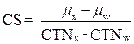
where X1
and X2 are the average mR/s values.
(m) The x-ray exposure control shall be so placed
that the operator can view the patient while making any exposure.
(n) Stationary x-ray systems, except veterinary
systems, shall be required to have the x-ray control permanently mounted in a
protected area that by virtue of shielding or distance or both shall protect
the operator during the entire exposure from radiation scatter and is
sufficient to comply with the occupational dose limits required by He-P
4020.05.
(o) Mobile and portable x-ray systems, except
veterinary systems shall meet the requirements of He-P 4046.06(n) if used
continuously for greater than one week in the same location,
and shall be provided with means to limit the source to skin distance
equal to, or greater than 30 centimeters.
(p) All stationary,
mobile or portable x-ray systems used for veterinary work shall be provided:
(1) With a 2 meter (6.5 feet) high protective
barrier for operator protection during exposures or
(2) With means to allow the operator to be at
least 2.7 meters (9 feet) from the tube housing assembly during the exposure.
(q) All mobile or portable radiographic systems,
except veterinary systems, shall be provided with means to limit the
source-to-skin distance to equal to or greater than 30 centimeters.
(r) When all technique factors are held
constant, including control panel selections associated with automatic exposure
control systems, the coefficient of variation of exposure for both manual and
automatic exposure control systems shall not exceed 0.05.
(s) Radiation emitted from the x-ray tube when
the system is fully charged and the exposure switch or timer is not activated
shall not exceed a rate of 2 milliroentgens per hour (mR/hr) at 5 centimeters from any accessible surface of the
diagnostic source assembly, with the beam-limiting device fully open.
(t) Deviation of measured technique factors
shall:
(1) Not exceed 10 percent of the indicated kVP value unless allowed by the manufacturer’s written
specification; and
(2) Not exceed 20 percent of the exposure time,
which is either indicated or which can be inferred from known mA or mAs values, unless allowed by the
manufacturer’s written specification.
(u) When equipment having independent selection
of x-ray tube current (mA), or a combined x-ray tube current exposure time
product (mAs), is operated between 40 percent and 100
percent of the maximum rated kVp, the absolute value
of the difference in the ratios (Xi) of exposure to the indicated
milliampere-seconds product mR/mAs
obtained at any two consecutive tube current settings shall not differ by more
than 0.10 times their sum:
where
the values of X1 and X2 are the average values which are
obtained at each of two consecutive tube current settings, or
are obtained at two settings differing by no more than a factor of 2 where the
tube current selection is continuous.
Source. #6827, eff 8-6-98; ss by #8692, INTERIM, eff
7-27-06, EXPIRES: 2-2-07; ss by #8808, eff 1-24-07; ss by #10763, INTERIM, eff
1-23-15, EXPIRES: 7-22-15; ss by #10893, eff 7-21-15 (from He-P 4046.07)
He-P 4046.07 Certified Diagnostic Systems Only. Diagnostic x-ray systems incorporating one or
more certified component(s) shall be required to comply with the following:
(a) For beam limitation for stationary
and mobile general purpose x-ray systems, the following shall be provided:
(1) A means of stepless adjustment of the size of
the x-ray field;
(2) A minimum field size at an
SID of 100 centimeters equal to or less than 5 centimeters by 5 centimeters;
(3) A light localizer, if used to define the
x-ray field, shall provide an average illumination, based upon measurements
made in the approximate center of each quadrant of the light field, of not less
than 160 lux or 15 foot-candles at 100 centimeters or the maximum SID,
whichever is less; and
(4) Radiation therapy simulation systems
manufactured on and after May 27, 1980, are exempt from the requirement of He-P
4046.07(a)(3);
(b) For beam limitation on stationary general
purpose x-ray systems equipped with positive beam limitation (PBL), the
following requirements shall be met:
(1) The PBL system shall be capable of operation,
at the discretion of the operator, such that the size of the field may be made
smaller than the size of the image receptor through stepless adjustment of the
field size;
(2) The minimum field size at an SID of 100
centimeters shall be equal to or less than 5 centimeters by 5 centimeters; and
(3) The PBL system shall function such that any
change of image receptor size or SID shall cause the automatic return to PBL;
(c) Beam limitation for portable x-ray systems
shall meet the beam limitation requirements of He‑P 4046.07(a) or (b) as
applicable;
(d) A tube stand or other mechanical support
shall be used for portable x-ray systems, so that the x-ray tube housing
assembly need not be hand-held during exposures; and
(e) Hand-held dental intraoral x-ray systems
shall not be subject to the requirements of He-P 4046.07(d).
Source. #6827, eff 8-6-98; ss by #8692, INTERIM, eff
7-27-06, EXPIRES: 2-2-07; ss by #8808, eff 1-24-07; ss by #10763, INTERIM, eff
1-23-15, EXPIRES: 7-22-15; ss by #10893, eff 7-21-15 (from He-P 4046.08)
He-P 4046.08 Dental Intraoral Radiographic Systems. In addition to the provisions of He-P 4045
and He-P 4046.01, the requirements of this section shall apply to x-ray systems
used for dental radiography:
(a) X-ray systems designed for use with an
intraoral image receptor shall be provided with means to limit SSD, to not less
than:
(1) 18 centimeters if operable above 50 kVp; or
(2) 10 centimeters if operable at 50 kVp only;
(b) X-ray systems designed for use with an
intraoral image receptor shall be provided with means to limit the x-ray beam
such that the beam at the minimum SSD shall be containable in a circle having a
diameter of no more than 7 centimeters;
(c) The radiation exposure control shall meet the
following requirements:
(1) No radiation exposure shall occur unless
initiated by the operator;
(2) It shall not be possible to make an exposure
when the timer is set to a “zero” or “off” position is provided;
(3) A visual indication of x-ray production shall
be observable by the operator;
(4) An audible signal to the operator shall
indicate that the exposure has terminated;
(5) Means shall be provided to terminate the
exposure at a preset time interval, preset product of current and time, a
preset number of pulses, or a preset radiation exposure to the image receptor;
(6) Exposures can be terminated by the operator
at any time, except for exposures of 0.5 second or less;
(7) Termination of an exposure shall cause
automatic resetting of the timer to its initial setting or to “zero”;
(8) Stationary intraoral systems shall be
required to have the x-ray exposure control permanently mounted in a protected
area, so that the operator is required to remain in that protected area during
the entire exposure;
(9) Mobile intraoral systems which are:
a. Used for greater than one week in the same
location, i.e., a room or suite, shall meet the requirements of He-P
4046.08(c)(8); or
b. Used for less than one week in the same
location shall be provided with either a protective barrier at least 2 meters
(6.5 feet) high for operator protection, or means to allow the operator to be
at least 2.7 meters (9 feet) from the tube housing assembly while making
exposures; and
(10) Hand-held dental intraoral x-ray machines
shall not be subject to the requirements of He-P 4046.08(c)(8) and He-P
4046.08(c)(9);
(d) When the equipment is operated on an
adequate power supply as specified by the manufacturer, the estimated
coefficient of variation of radiation exposures shall be no greater than 0.05,
for any specific combination of selected technique factors;
(e) Deviation of technique factors from indicated
values for kVp and exposure time shall not exceed the
limits specified for that system by its manufacturer or in the absence of
manufacturer’s specifications the deviation shall not exceed 10 percent of the
indicated value for kVp and 20 percent for time;
(f) Dental x-ray systems with a nominal fixed kVp of less than 50 kVp shall not
be used to make diagnostic dental radiographs of humans; and
(g) The following administrative controls shall
be in place:
(1) Patient and image receptor holding devices
shall be used when the techniques permit;
(2) Neither the tube housing nor any position
indicating device (PID) such as a Rinn kit shall be hand-held during an
exposure; and
(3) Dental fluoroscopy without image
intensification shall not be used.
Source. #6827, eff 8-6-98; ss by #8692, INTERIM, eff
7-27-06, EXPIRES: 2-2-07; ss by #8808, eff 1-24-07; ss by #10763, INTERIM, eff
1-23-15, EXPIRES: 7-22-15; ss by #10893, eff 7-21-15 (from He-P 4046.09)
He-P 4046.09 Hand-held Dental Intraoral Machines. The following requirements shall apply to the
use of hand-held dental intraoral machines:
(a) For all uses:
(1) The registrant shall follow all applicable
requirements set forth in the New Hampshire Rules for the Control of Radiation
(NHRCR), as well as all manufacturers’s
requirements;
(2) The machine shall be used only for dental
diagnostic imaging;
(3) The protective shielding shall not be removed
or modified;
(4) The registrant shall provide a copy of their
operating, safety and security procedures to operators to prevent unauthorized
or improper use;
(5) The registrant shall prepare a quality
assurance plan, with an emphasis on ensuring limited retaking of radiographs;
(6) Operators of hand-held dental intraoral
machines shall be specifically trained to operate such equipment, and the
registrant shall provide proof of training for all operators;
(7) When operating a hand-held dental intraoral
machine, operators shall wear a lead apron as well as
an extremity dosimeter on the hand closest to the protective shielding;
(8) A hand-held dental intraoral machine shall be
held without any motion during a patient examination. A tube stand or Rinn kit may be utilized to
immobilize a hand-held dental intraoral radiographic unit during patient
examination;
(9) Operators of hand-held dental intraoral
machines shall provide a secondary protective barrier for patients; and
(10) The operator shall ensure that there are no
bystanders within a radius of at least 6 feet (2 meters), from the patient
being examined with a hand-held intraoral machine.
(b) Additional requirements for operatories in
permanent facilities:
(1) Hand-held dental intraoral machines shall be
used for patient examinations only in dental operatories that meet the
structural shielding requirements specified by DHHS/RHS or by a qualified
expert;
(2) Hand-held dental intraoral machines shall not
be used for patient examinations in hallways and waiting rooms; and
(3) For battery-powered units, visual means shall
be provided on the control panel to indicate whether the battery is in a state
of charge adequate for proper operation.
Source. #6827, eff 8-6-98; ss by #8692, INTERIM, eff
7-27-06, EXPIRES: 2-2-07; ss by #8808, eff 1-24-07; ss by #10763, INTERIM, eff
1-23-15, EXPIRES: 7-22-15; ss by #10893, eff 7-21-15
He-P 4046.10 Dental Cone-Beam CT (CBCT). Dental systems shall
be:
(a) Certified by the manufacturer pursuant to the
Medical Device Act and Subchapter C-Electronic Product Radiation Control (EPRC)
of Chapter V of the Federal Food, Drug and Cosmetic Act;
(b) Registered in accordance with He-P 4040;
(c) Maintained and operated in accordance with
the manufacturer’s specification; and
(d) Operated by persons
who have been specifically trained by the manufacturer.
Source. #6827, eff 8-6-98; ss byD,
INTERIM, eff 7-27-06, EXPIRES: 2-2-07; ss by #8808, eff 1-24-07; ss by #10763, INTERIM,
eff 1-23-15, EXPIRES: 7-22-15; ss by #10893, eff 7-21-15
He-P 4046.11 Computed Tomography X-ray System
Requirements.
(a) Automatic termination of x-ray exposures in
the event of equipment failure shall occur either by de-energizing the x-ray
source or shuttering the x-ray beam.
(b) Termination as required in He-P 4046.11(a)
above shall occur within an interval that limits the total scan time to no more
than 110 percent of its preset value.
(c) Premature termination of the x-ray exposure
by the operator shall require resetting of the CT conditions of operation prior
to initiation of another scan.
(d) A visible signal shall indicate when the
x-ray exposure has been terminated.
(e) The operator shall be able to terminate the
x-ray exposure at any time during a scan of greater than one-half second
duration.
(f) For any single tomogram system, means shall
be provided to permit visual determination of the tomographic plane or a
reference plane offset from the tomographic plane.
(g) For any multiple tomogram system, means shall
be provided to permit visual determination of the location of a reference
plane.
(h) If a device using a light source is used to
satisfy the requirements of He-P 4046.11(f) or (g) above, the light source
shall provide illumination levels sufficient to permit visual determination of
the location of the tomographic plane or reference plane under ambient light
conditions of up to 500 lux.
(i) The CT x-ray
control and gantry shall provide visual indication whenever x-rays are produced
and, if applicable, whether the shutter is open or closed.
(j) Each emergency button or switch shall be
clearly labeled as to its function.
(k) The CT conditions of operation to be used
during a scan or a scan sequence shall be indicated prior to the initiation of
a scan or a scan sequence.
(l) Indication of CT conditions of operation
shall be visible from any position from which scan initiation is possible.
(m) The angular position where the maximum
surface CTDI occurs shall be identified to allow for reproducible positioning
of a CT dosimetry phantom.
(n) Additional requirements applicable to CT
X-ray systems containing a gantry manufactured after September 3, 1985 shall be as follows:
(1) The total error in the indicated location of
the tomographic plane or reference plane shall not exceed 5 millimeters;
(2) If the x-ray production period is less than
one-half second, the indication of x-ray production shall be actuated for at
least one-half second;
(3) Indicators at or near the gantry shall be
discernible from any point external to the patient opening;
(4) The deviation of indicated scan increment
versus actual increment shall not exceed plus or minus one millimeter with any
mass from 0 to 100 kilograms resting on the support device; and
(5) The patient support device shall be
incremented from a typical starting position to the maximum incremented
distance or 30 centimeters, whichever is less, and then returned to the
starting position. Measurement of actual versus indicated scan increment may be
taken anywhere along the incremented distance.
Source. #6827, eff 8-6-98; ss by #8692, INTERIM, eff
7-27-06, EXPIRES: 2-2-07; ss by #8808, eff 1-24-07; ss by #10763, INTERIM, eff
1-23-15, EXPIRES: 7-22-15; ss by #10893, eff 7-21-15 (from He-P 4046.11)
He-P 4046.12 Computed Tomography X-ray Systems –
Facility Design Requirements.
(a) Provision shall be made for 2-way aural
communication between the patient and the operator at the control panel.
(b) Windows, mirrors, closed-circuit television,
or an equivalent shall be provided to permit continuous observation of the
patient during irradiation and shall be so located that the operator can
observe the patient from the control panel.
(c) When the primary viewing system is by
electronic means, an alternate viewing system shall be available for use in the
event of failure of the primary viewing system.
Source. #6827, eff 8-6-98; ss by #8692, INTERIM, eff
7-27-06, EXPIRES: 2-2-07; ss by #8808, eff 1-24-07; ss by #10763, INTERIM, eff
1-23-15, EXPIRES: 7-22-15; ss by #10893, eff 7-21-15 (from He-P 4046.11)
He-P 4046.13 Computed Tomography X-ray Systems –
Surveys, Calibrations, Spot Checks, and Operating Procedures.
(a) All CT x-ray systems shall have a survey made by or under the direction of a qualified expert.
(b) The registrant shall obtain a written report
of the survey from the qualified expert, and a copy of the report shall be made
available to DHHS/RHS upon request.
(c) The calibration of the radiation output of
the CT x-ray system shall be performed by or under the direction of a qualified
expert who is physically present at the facility during such calibration.
(d) The calibration of a CT x-ray system shall be
performed at intervals specified by a qualified expert and after any change or
replacement of components which, in the opinion of the qualified expert, could
cause a change in the radiation output.
(e) The calibration of the radiation output of a
CT x-ray system shall be performed with a calibrated dosimetry system which is
traceable to a national standard and has been calibrated within the preceding 2
years.
(f) CT dosimetry phantom(s) shall meet the
following specifications and conditions of use shall be used in determining the
radiation output of a CT X-ray system:
(1) CT dosimetry phantom(s) shall be right
circular cylinders of polymethyl methacrylate of density 1.19 plus or minus
0.01 grams per cubic centimeter;
(2) The phantom(s) shall be at least 14
centimeters in length and shall have diameters of 32.0 centimeters for testing
CT x-ray systems designed to image any section of the body and 16.0 centimeters
for systems designed to image the head or for whole body scanners operated in
the head scanning mode;
(3) CT dosimetry phantom(s)
shall provide means for the placement of a dosimeter(s) along the axis
of rotation and along a line parallel to the axis of rotation 1.0 centimeter
from the outer surface and within the phantom; and
(4) All dose measurements shall be performed with
the CT dosimetry phantom placed on the patient couch or support device without
additional attenuation materials present.
(g) The calibration shall be required for each
type of head, body, or whole-body scan performed at the facility.
(h) Calibration shall meet the following
requirements:
(1) The dose profile along the center axis of the
CT dosimetry phantom for the minimum, maximum, and midrange values of the
nominal tomographic section thickness used by the registrant shall be
measurable;
(2) Where less than 3 nominal tomographic
thicknesses can be selected, the dose profile determination shall be performed
for each available nominal tomographic section thickness;
(3) The CTDI along the two axes specified in He-P
4046.02 (l)(1) shall be measured;
(4) The CT dosimetry phantom shall be oriented so
that the measurement point 1.0centimeter from the
outer surface and within the phantom is in the same angular position within the
gantry as the point of maximum surface CTDI identified; and
(5) The CT conditions of operation shall
correspond to typical values used by the registrant for body parts represented
by that phantom type.
(i) Calibration
procedures shall be in writing.
(j) Records of calibrations performed shall be
maintained for inspection by DHHS/RHS.
(k) The spot check procedures shall be in writing
and shall have been developed by a qualified expert.
(l) The spot check procedures shall incorporate
the use of a CT dosimetry phantom capable of providing an indication of
contrast scale, noise, nominal tomographic section thickness, resolution
capability of the system for low and high contrast objects,
and measuring the mean CTN for water or other reference material.
(m) All spot checks shall be included in the CT
calibration and at time intervals and under system conditions specified by a
qualified expert.
(n) Spot checks shall include acquisition of
images obtained with the CT dosimetry phantom(s) using the same processing mode
and CT conditions of operation as are used to perform calibrations.
(o) The spot check images shall be retained,
until a new calibration is performed, as images stored in digital form on a
backed-up storage medium compatible with the CT x-ray system.
(p) Written records of the spot checks performed
shall be maintained for inspection by DHHS/RHS.
(q) The CT x-ray system shall not be operated
except by a person who has been specifically trained in its operation.
(r) Information shall be available at the control
panel regarding the operation and calibration of the system to include the
following:
(1) Dates of the latest calibration and
spot-checks and the location within the facility where the results of those
tests may be obtained;
(2) Instructions on the use of the CT dosimetry
phantom(s) including a schedule of spot-checks appropriate for the system,
allowable variations for the indicated parameters, and the results of at least
the most recent spot-checks conducted on the system;
(3) The distance in millimeters between the
tomographic plane and the reference plane if a reference plane is utilized;
(4) A current technique chart available at the
control panel which specifies for each routine examination the CT conditions of
operation and the number of scans per examination; and
(5) If the calibration or spot- check of the CT
x-ray system identifies that a system operating parameter has exceeded a
tolerance established by the qualified expert, use of the CT x-ray system on
patients shall be limited to those uses permitted by established written
instructions of the qualified expert.
Source. #6827, eff 8-6-98; ss by #8692, INTERIM, eff
7-27-06, EXPIRES: 2-2-07; ss by #8808, eff 1-24-07; ss by #10763, INTERIM, eff
1-23-15, EXPIRES: 7-22-15; ss by #10893,
eff 7-21-15 (from He-P 4046.12)
He-P 4046.14 Mammography X-ray System Requirements.
(a) In addition to other rules pertaining to
radiation safety, registration and use of radiation machines, only radiation
machines pursuant to the “Mammography Quality Standards Reauthorization Act of
1998,” Public Law 105-248, and 21 CFR Part 900, shall be used for screening and
diagnostic mammography.
(b) A facility performing screening and
diagnostic mammography shall have a valid certificate issued by the US
Department of Health and Human Services, pursuant to the “Mammography Quality
Standards Reauthorization Act of 1998,” Public Law 105-248, and 21 CFR Part
900.
Source. #8808, eff 1-24-07; ss by #10763, INTERIM,
eff 1-23-15, EXPIRES: 7-22-15; ss by #10893, eff 7-21-15 (from He-P 4046.13)
He-P 4046.15 Bone Densitometry.
(a) When an application for registration of a new
or additional bone densitometry machines is received
by DHHS/RHS in accordance with He-P 4040, registration shall be denied unless
the application is accompanied by the following information:
(1) A detailed description of the x-ray
examinations proposed in the screening program;
(2) A description of the diagnostic x-ray quality
control program;
(3) The qualifications of each person who will be
operating the x-ray system(s);
(4) The qualifications of the person who will be
supervising the operators of the x-ray systems(s), the extent of supervision,
and the method of work performance evaluation; and
(5) The name and address of the person who will
interpret the radiograph(s).
(b) Bone densitometry systems shall be:
(1) Certified by the manufacturer pursuant to the
Medical Device Act and Subchapter C- Electronic Product Radiation Control
(EPRC) of Chapter V of the Federal Food, Drug and Cosmetic Act;
(2) Licensed in accordance with He-P 4040 of
these regulations; and
(3) Maintained and operated in accordance with
the manufacturer’s specifications.
(c) Bone densitometry systems with a stepless
beam limitation device shall be provided with means to both size and align the
x-ray field such that at a plane of the image receptor the x-ray field does not
extend beyond 2 percent of the SID.
(d) Operators of bone densitometry systems shall
be:
(1) Licensed as a practitioner of the healing arts; or
(2) Complete a training course on bone
densitometry which is provided by the densitometer manufacturer or vendor, and
which shall include:
a. Basic radiation protection;
b. Operating procedures for bone densitometry
systems, to include use of various system functions, safety and maintenance;
and
c. Patient positioning for the types of
examinations performed.
(e) During the operation of any bone densitometry
system:
(1) The operator, ancillary personnel, and
members of the general public shall be positioned at
least one meter from the patient and bone densitometry system during the
examination; and
(2) The operator shall
advise the patient that the bone densitometry examination is a type of x-ray
procedure.
(f) The registrant shall keep maintenance records
for bone densitometry records as prescribed by He-P 4045.05. These records shall be maintained for
inspection DHHS/RHS for a minimum period of 5 years.
(g) Bone densitometry on human patients shall be
conducted only under the prescription of a licensed practitioner of the healing
arts.
Source. #10893, eff 7-21-15 (from He-P 4046.14)
He-P 4046.16 Veterinarian Facilities – Radiation or MRI
Machine General Requirements.
(a) All veterinarian facilities shall be subject
to the following requirements:
(1) Radiation machine general requirements set
forth in He-P 4046.01; and
(2) Tube potential vs., minimum half-value layer
set forth in Table 4046.1.
(b) Veterinarians using fluoroscopic systems
shall be subject to requirements for:
(1) General requirements and activation of the fluoro tube set forth in He-P 4046.02;
(2) Barrier transmitted exposure rate limits set
forth in He-P 4046.04;
(3) Additional requirements set forth in He-P
4046.05; and
(4) Certified diagnostic systems set forth in
He-P 4046.07.
(c) Veterinarians using dental intraoral systems
shall be subject to the requirements for:
(1) Dental intraoral radiographic systems, other
than hand-held systems set forth in He-P 4046.08; and
(2) Hand-held dental intraoral systems set forth
in He-P 4046.09.
(d) Veterinarians using computed tomography
systems shall be subject to requirements for:
(1) Equipment requirements set forth in He-P
4046.11;
(2) Facility design requirements set forth in
He-P 4046.12; and
(3) Surveys, calibrations, spot checks, and
operating conditions set forth in He-P 4046.13.
Source. #10893, eff 7-21-15
He-P 4046.17 Magnetic Resonance Imaging (MRI) Systems. MRI systems shall be:
(a) Certified by the manufacturer pursuant to the
Medical Device Act and Subchapter C-Electronic Product Radiation Control (EPRC)
of Chapter V of the Federal Food, Drug and Cosmetic Act;
(b) Registered in accordance with He-P 4040; and
(c) Maintained and operated in accordance with
the manufacturer’s specifications.
Source. #10893, eff 7-21-15
PART He-P 4047 USE OF RADIATION
MACHINES: THERAPEUTIC RADIATION MACHINES
He-P
4047.01 Purpose. He-P 4047 establishes
requirements, for which the registrant is responsible for use of therapeutic
radiation machines.
Source. #6827, eff 8-6-98; ss by #8692, INTERIM, eff
7-27-06, EXPIRES: 2-2-07; ss by #8808, eff 1-24-07; ss by #10764, INTERIM, eff
1-23-15, EXPIRES: 7-22-15; ss by #10894, eff 7-21-15
New. #14598, INTERIM, eff 5-22-26, EXPIRES:
11-18-26
He-P
4047.02 Scope. The provisions of He-P 4047 are in
addition to, and are not substitutions for, other applicable provisions of this
chapter.
Source. #6827, eff 8-6-98; ss by #8692, INTERIM, eff
7-27-06, EXPIRES: 2-2-07; ss by #8808, eff 1-24-07; ss by #10764, INTERIM, eff
1-23-15, EXPIRES: 7-22-15; ss by #10894, eff 7-21-15
New. #14598, INTERIM, eff 5-22-26, EXPIRES:
11-18-26
He-P
4047.03 Definitions. The following definitions
apply specifically to Part He-P 4047 and shall be in addition to definitions in
He-P 4003 and He-P 4041:
(a) “Absorbed
dose (D)” means the mean energy imparted by ionizing radiation to matter, and is determined as the quotient of dE by dm, where dE is
the mean energy imparted by ionizing radiation to matter of mass dm, and is
expressed in the SI unit of joule per kilogram and the special name “gray”
(Gy).
(b) “Absorbed
dose rate” means absorbed dose per unit time for radiation machines with
timers, or dose monitor unit per unit time for linear accelerators.
(c) “Air kerma (K)” means the kinetic energy released in air by
ionizing radiation, and is determined as the quotient
of dE by dM,
where dE is the sum of the initial kinetic
energies of all the charged ionizing particles liberated by uncharged ionizing
particles in air of mass dM, and is expressed in
the SI unit of joule per kilogram, and the special name the “gray” (Gy).
(d) “Beam
scattering foil” means a thin piece of material placed in the beam to scatter a
beam of electrons in order to provide a more uniform
electron distribution in the useful beam.
(e) “Bent
beam linear accelerator” means a linear accelerator geometry in which the
accelerated electron beam must change direction by passing through a bending
magnet.
(f) “Conventional
simulator” means any x-ray system designed to produce the geometric conditions
of the radiation therapy equipment.
(g) “Dose
monitor unit (DMU)” means a unit response from the beam monitoring system from
which the absorbed dose can be calculated.
(h) “Dosimetrist”
means a person who generates and calculates radiation dose distributions under
the direction of a medical physicist and a radiation oncologist.
(i) “Electronic brachytherapy” means a method of
radiation therapy where an electrically generated source of ionizing radiation
is placed in or near the tumor or target tissue to deliver therapeutic
radiation dosage.
(j) “Electronic
brachytherapy device” means the system used to produce and deliver therapeutic
radiation including the x-ray tube, the control mechanism, the cooling system,
and the power source.
(k) “Electronic
brachytherapy source” means the x-ray tube component used in an electronic
brachytherapy device.
(l) “External
beam radiation therapy” means therapeutic irradiation in which the source of
radiation is at a distance from the body.
(m) “Field-flattening
filter” means a filter used to homogenize the absorbed dose rate over the
radiation field.
(n) “Gantry”
means that part of a radiation therapy system supporting and allowing movements
of the radiation head about a center of rotation.
(o) “High
energy external beam therapy” means photons and electrons with energies greater
than equal to one MV or one MeV.
(p) “Intensity
Modulated Radiation Therapy (IMRT)” means radiation therapy that uses
non-uniform radiation beam intensities which have been determined by various
computer based and automated optimization techniques.
(q) “Interruption
of irradiation” means the stopping of irradiation with the possibility of
continuing irradiation without resetting of operating conditions at the control
panel.
(r) “Kilovolt
(kV)” or “kilo electron volt (keV)” means the energy equal to that acquired by
a particle with one electron charge in passing through a potential difference
of one thousand volts in a vacuum.
(s) “Megavolt
(MV)” or “mega electron volt (MeV)” means the energy equal to that acquired by
a particle with one electron charge in passing through a potential difference
of one million volts in a vacuum.
(t) “Misadministration”
means the administration of an external beam radiation therapy dose:
(1) Involving
the wrong patient;
(2) Involving
the wrong treatment modality;
(3) Involving
the wrong treatment site;
(4) When
the treatment consists of 3 or fewer fractions and the calculated total
administered dose differs from the total prescribed dose by more than 10
percent of the total prescribed dose;
(5) When
the calculated weekly administered dose differs from the weekly prescribed dose
by more than 30 percent; or
(6) When
the calculated total administered dose differs from the total prescribed dose
by more than 20 percent of the total prescribed dose.
(u) “Mobile
Electronic Brachytherapy Service” means transportation of an electronic
brachytherapy device to provide electronic brachytherapy at an address that is
not the address of record.
(v) “Monitor
unit (MU)” means “dose monitor unit” as defined in (g) above.
(w) “Moving
beam radiation therapy” means radiation therapy with any planned displacement
of radiation field or patient relative to each other, or with any planned
change of absorbed dose distribution.
(x) “Nominal
treatment distance” means:
(1) For
electron radiation, the distance from the scattering foil, virtual source, or
exit window of the electron beam to the entrance surface of the irradiation
object along the central axis of the useful beam;
(2) For
x-ray irradiation, the virtual source or target to isocenter distance along the
central axis of the useful beam; and
(3) For
non-isocentric x-ray irradiation equipment, the
distance along the central axis shall be that specified by the manufacturer.
(y) “Periodic
quality assurance check” means a procedure which is performed to ensure that a
previous calibration continues to be valid.
(z) “Phantom”
means an object behaving in essentially the same manner as tissue, with respect
to absorption or scattering of the ionizing radiation in question.
(aa) “Practical
range of electrons” means a classical electron range where the only remaining
contribution to dose is from bremsstrahlung x-rays.
(ab) “Prescribed
dose” means the total dose and dose per fraction as documented in the written
directive.
(ac) “Primary
dose monitoring system” means a system which will monitor the useful beam
during irradiation and which will terminate
irradiation when a pre-selected number of dose monitor units have been
delivered.
(ad) “Radiation
field” means “useful beam” as defined in He-P 4041(fn).
(ae) “Radiation
head” means the structure from which the useful beam emerges.
(af) “Radiation therapy physicist” means an
individual qualified in accordance with He‑P 4047.
(ag) “Recordable
event” means the administration of an external beam radiation therapy dose when
the calculated weekly administered dose differs by 15 percent or more from the
weekly prescribed dose.
(ah) “Redundant
beam monitoring system” means a combination of 2 dose monitoring systems in
which each system is designed to terminate irradiation in accordance with a
pre-selected number of dose monitor units.
(ai) “Scattered
primary radiation” means that scattered radiation which has been deviated in
direction only by materials irradiated by the useful beam.
(aj) “Secondary dose monitoring system” means a
system which will terminate irradiation in the event of failure of the primary
dose monitoring system.
(ak) “Shadow tray” means a device attached to the
radiation head to support auxiliary beam blocking material.
(al) “Simulator”
or “radiation therapy simulation system” means an x-ray system intended for
localizing the volume to be exposed during radiation therapy and reproducing
the position and size of the therapeutic irradiation field.
(am) “Source-skin
distance (SSD)” means “target-skin distance” as defined in (ap) below.
(an) “Stationary
beam radiation therapy” means radiation therapy without displacement of one or
more mechanical axes relative to the patient during irradiation.
(ao) “Target” means that part of an x-ray tube or
accelerator onto which is directed a beam of accelerated particles to produce
ionizing radiation or other particles.
(ap) “Target-skin
distance (TSD)” means the distance measured along the beam axis from the center
of the front surface of the x-ray target or electron virtual source to the
surface of the irradiated object or patient.
(aq) “Therapeutic radiation machine” means x-ray
or electron producing equipment designed and used for external beam radiation
therapy. For the purpose of these
regulations, devices used to administer electronic brachytherapy shall also be
considered therapeutic machines.
(ar) “Virtual Simulator” means a computed
tomography (CT) unit used in conjunction with relevant software which recreates
the treatment machine; and that allows import manipulation, display and storage
of images from CT or other imaging modalities.
(as) “Virtual
source” means a point from which radiation appears to originate.
(at) “Written
directive” means an order in writing for a specific patient, dated and signed
by an authorized user prior to the administration of radiation which contains
the total dose, dose per fraction, treatment site, and overall treatment period.
Source. #6827, eff 8-6-98; ss by #8692, INTERIM, eff
7-27-06, EXPIRES: 2-2-07; ss by #8808, eff 1-24-07; ss by #10764, INTERIM, eff
1-23-15, EXPIRES: 7-22-15; ss by #10894, eff 7-21-15
New. #14598, INTERIM, eff 5-22-26, EXPIRES:
11-18-26
He-P 4047.04 General Administrative Requirements for
Facilities Using Therapeutic Radiation Machines.
(a) The
use of therapeutic radiation machines shall be by, or under the supervision of,
a licensed practitioner of the healing arts.
(b) The
registrant shall be responsible for directing the operation of the therapeutic
radiation machines which have been registered with DHHS/RHS.
(c) The
registrant shall ensure that the requirements of He-P 4047 are met in the
operation of the therapeutic radiation machine(s).
(d) A
therapeutic radiation machine which does not meet the provisions of this part
shall not be used for irradiation of patients.
(e) The
registrant for any therapeutic radiation machine subject to He-P 4047 shall
require the authorized user to be a physician who:
(1) Is
certified in:
a. Radiation
oncology or therapeutic radiology by the American Board of Radiology;
b. Radiation
oncology by the American Osteopathic Board of Radiology;
c. Radiology,
with specialization in radiotherapy, as a British “Fellow of the Faculty of
Radiology” or “Fellow of the Royal College of Radiology”; or
d. Therapeutic
radiology by the Canadian Royal College of Physicians and Surgeons; or
(2) Is
in the active practice of therapeutic radiology, and has:
a. Completed
200 hours of instruction in basic radiation techniques applicable to the use of
an external beam radiation therapy unit;
b. Completed
500 hours of supervised work experience; and
c. A
minimum of 3 years supervised clinical experience.
(f) To
satisfy the requirement for instruction in (e)(2)a. above, the classroom and
laboratory training shall include:
(1) Radiation
physics and instrumentation;
(2) Radiation
protection;
(3) Mathematics
pertaining to the use and measurement of ionization radiation; and
(4) Radiation
biology.
(g) To
satisfy the requirement for supervised work experience in (e)(2)b. above,
training shall:
(1) Be
under the supervision of an authorized user; and
(2) Include:
a. Review
of the full calibration measurements and periodic quality assurance checks;
b. Evaluation
of prepared treatment plans;
c. Calculation
of treatment times and patient treatment settings;
d. Use
of administrative controls to prevent misadministrations;
e. Implementation
of emergency procedures to be followed in the event of the abnormal operation
of a external beam
radiation therapy unit or console; and
f. The
checking and use of radiation survey meters.
(h) To satisfy the
requirement for a period of supervised clinical experience in (e)(2)c. above,
training shall include:
(1) One
year in a formal training program approved by:
a. The
Residency Review Committee for Radiology of the Accreditation Council for
Graduate Medical Education; or
b. The
Committee on Postdoctoral Training of the American Osteopathic Association;
(2) An
additional 2 years of clinical experience in therapeutic radiology under the
supervision of an authorized user; and
(3) The
following training:
a. Examining
individuals;
b. Reviewing
individuals’ case histories to determine their suitability for external beam
radiation therapy treatment, and any limitations and contraindications;
c. Selecting
proper dose;
d. Selecting
how the dose is to be administered;
e. Calculating
the external beam radiation therapy doses;
f. Collaborating
with the authorized user in the review of patients’ progress;
g. Considering of the need to modify originally prescribed
doses or treatment plans as warranted by patients’ reaction to radiation;
h. Post-administration
follow-up; and
i. Post-administration
review of case histories.
(i) Notwithstanding the requirements of (e)(1)
and (e)(2) above, the registrant for any therapeutic radiation machine subject
to He-P 4047 shall also submit the training of the prospective authorized user
physician for DHHS/RHS review.
(j) A
physician shall not act as an authorized user for any therapeutic radiation
machine until such time as said physician’s training has been reviewed and
approved by DHHS/RHS.
(k) The
registrant for any therapeutic radiation machine subject to He-P 4047 shall
require the radiation therapy physicist to be registered with DHHS/RHS, under
the provisions of He-P 4040, as a provider of radiation services in the area of
calibration and compliance surveys of external beam radiation therapy units,
and either:
(1) Be
certified by:
a. The
American Board of Radiology in:
1. Therapeutic radiological
physics;
2. Roentgen-ray and gamma-ray
physics;
3. X-ray and radium physics; or
4. Radiological physics;
b. The
American Board of Medical Physics in Radiation Oncology Physics; or
c. The
Canadian College of Medical Physics; or
(2) Alternately,
the radiation therapy physicist shall meet the following requirements:
a. Hold
a master’s or doctor’s degree in physics, medical physics, engineering, applied
mathematics, biophysics, radiological physics, or health physics from an
accredited college or university;
b. Have
completed one year of full time training in
therapeutic radiological physics;
c. Have
completed one year full-time work experience under the
supervision of a radiation therapy physicist at a medical institution; and
d. The
training and work experience as required in b. and c.
above, shall be conducted in a clinical radiation facility that provides
high-energy external beam radiation therapy.
(l) To
meet the requirement in (k)(2)c. above, the individual shall have performed the
tasks listed in He-P 4047.05(a) through (f), and He-P 4047.06(r) and (s), and
He-P 4047.07(u) and (v), under the supervision of a radiation therapy physicist
during the year of work experience.
(m) Notwithstanding
the provisions of (k)(2) and (l) above, certification pursuant to (k)(1) above
shall be required on or before December 31, 1999 for
all persons currently qualifying as a radiation
therapy physicist pursuant to (k) above.
(n) Each
individual who operates a therapeutic radiation machine for medical use shall:
(1) Be
a registered radiation therapy technologist with the American Registry of
Radiologic Technologists (ARRT);
(2) Submit
evidence that they have satisfactorily completed a radiation therapy
technologist training program that complies with the requirements of the Joint
Review Committee on Education in Radiologic Technology; or
(3) Be
a student working under the direct supervision and in the physical presence of
a registered radiation therapy technologist.
(o) The
names and training of all personnel currently operating a therapeutic radiation
machine shall be kept on file at the facility.
(p) Information
about former operators shall be retained for a period of at least 2 years
beyond the last date they were authorized to operate a therapeutic radiation
machine at that facility.
(q) Written
safety procedures and rules shall be developed by a radiation therapy
physicist.
(r) Written
safety procedures and rules shall be available in the control area of a
therapeutic radiation machine, including any restrictions required for the safe
operation of the particular therapeutic radiation
machine.
(s) The
operator shall be able to demonstrate familiarity with the procedures and rules
required in (q) and (r) above.
(t) Individuals shall not be
exposed to the useful beam except for medical therapy purposes and unless such
exposure has been ordered in writing by an authorized user.
(u) The
provision in (t) above shall specifically prohibit deliberate exposure of an
individual for training, demonstration, or other non-healing-arts purposes.
(v) Notwithstanding
the provisions of (e)(1) and (e)(2) above, a registrant may permit any
physician to act as a visiting authorized user under the term
of the registrant’s Certificate of Registration for up to 60 days per calendar year under the following conditions:
(1) The
visiting authorized user has the prior written permission of the registrant’s
management;
(2) The
use occurs on behalf of an institution, or the institution’s radiation safety
committee;
(3) The
visiting authorized user meets the requirements established for authorized
user(s) in (e)(1) and (e)(2) above; and
(4) The
registrant maintains copies of all records specified by the paragraph for 5
years after the records are made.
(w) All
individuals associated with the operation of a therapeutic radiation machine
shall be instructed in the provisions of the registrant’s quality management
program.
(x) All
individuals associated with the operation of a therapeutic radiation machine
shall comply with the provisions of the registrant’s quality management
program.
(y) All
individuals associated with the operation of a therapeutic radiation machine
shall be subject to the requirements of He-P 4020 through He-P 4022.
(z) The
registrant shall maintain for inspection by DHHS/RHS the following information
in a separate file or package for each therapeutic radiation machine:
(1) Report
of acceptance testing;
(2) Records
of all:
a. Surveys
with date(s) performed;
b. Calibrations
with date(s) performed;
c. Periodic,
dated, quality assurance checks of the therapeutic radiation machine required
by He-P 4047; and
d. The
name(s) of the person(s) who performed such activities;
(3) Records
of:
a. Maintenance,
with the date(s) it was performed;
b. Any
modifications, with the date(s) the modifications were performed on the
therapeutic radiation machine; and
c. The
name(s) of the person(s) who performed such services; and
(4) The
signature of the person authorizing the return of therapeutic radiation machine
to clinical use after service, repair, or upgrade.
(aa) All
records required by He-P 4047 shall be retained until disposal is authorized by
DHHS/RHS unless another retention period is specifically authorized in He-P
4047.
(ab) All
required records shall be retained in an active file from at least the time of
generation until the next DHHS/RHS inspection.
(ac) Any
required record generated prior to the last DHHS/RHS inspection may be
microfilmed or otherwise archived as long as a complete copy of said record can
be retrieved until such time as DHHS/RHS authorizes final disposal.
Source. #6827, eff 8-6-98; ss by #8692, INTERIM, eff
7-27-06, EXPIRES: 2-2-07; ss by #8808, eff 1-24-07; ss by #10764, INTERIM, eff
1-23-15, EXPIRES: 7-22-15; ss by #10894, eff 7-21-15
New. #14598, INTERIM, eff 5-22-26, EXPIRES:
11-18-26
He-P 4047.05 General Technical Requirements for
Facilities Using Therapeutic Radiation Machines.
(a) The
registrant shall ensure that radiation protection surveys of all new
facilities, and existing facilities not previously surveyed, are performed with
an operable radiation measurement survey instrument calibrated in accordance
with He-P 4047.08.
(b) The
radiation protection survey shall be performed by, or under the direction of, a
radiation therapy physicist or a qualified expert.
(c) The
radiation protection survey shall verify that, with the therapeutic radiation
machine in a “BEAM-ON” condition, with the largest clinically available
treatment field and with a scattering phantom in the useful beam of radiation,
the following requirements are met:
(1) Radiation
levels in restricted areas are not likely to cause personnel exposures in excess of the limits specified in He-P 4020.05; and
(2) Radiation
levels in unrestricted areas do not exceed the limits specified in He-P
4020.13(a) and (b).
(d) In
addition to the requirements of (a) above, a radiation protection survey shall
also be performed prior to any subsequent medical use;
and:
(1) After
making any change in the treatment room shielding;
(2) After
making any change in the location of the therapeutic radiation machine within
the treatment room;
(3) After
relocating the therapeutic radiation machine to a different treatment room; or
(4) Before
using the therapeutic radiation machine in a manner that could result in
increased radiation levels in areas outside the external radiation therapy
treatment room.
(e) The
survey record required in (d) above shall include:
(1) All
instances where the facility is in violation of applicable rules;
(2) The
date of the measurements;
(3) The
reason for the survey;
(4) The
manufacturer’s name;
(5) Model
number of the therapeutic radiation machine;
(6) Serial
number of the therapeutic radiation machine;
(7) The
instrument(s) used to measure radiation levels;
(8) A
diagram of the areas surrounding the treatment room that were surveyed;
(9) The
measured dose rate at several points in each area expressed in microsieverts or millirems per hour;
(10) The
calculated maximum level of radiation over a period of 1 week for each
restricted and unrestricted area; and
(11) The
signature of the individual responsible for conducting the survey.
(f) If
the results of the surveys required by (a) and (d) above indicate any radiation
levels in excess of the respective limit specified in He-P 4020, the registrant
shall lock the control in the “OFF” position and not use the unit except as might
be necessary to repair, replace, or test the therapeutic radiation machine,
test the therapeutic radiation machine shielding, or test the treatment room
shielding.
(g) If
the survey required by (a) and (d) above indicates that an individual in an
unrestricted area might be exposed to levels of radiation greater than those
permitted by He-P 4020.13(a) and (b), before beginning the treatment program
the registrant shall:
(1) Equip
the unit with beam direction interlocks or add additional radiation shielding
to ensure compliance with He-P 4020.13(a) and (b);
(2) Perform
the survey required by (a) above again; and
(3) Include
in the record required by (e) above:
a. The
results of the initial survey;
b. A
description of the modification made to comply with (g)(1) above; and
c. The
results of the second survey.
(h) The
registrant shall have a dosimetry system available for use which has been
calibrated by the National Institute for Standards and Technology (NIST) or by
an American Association of Physicists in Medicine (AAPM) Accredited Dosimetry
Calibration Laboratory (ADCL) as follows:
(1) For
beams with energies greater than 1 MV (1 MeV), the dosimetry system shall have
been calibrated for Cobalt-60; and
(2) For
beams with energies equal to or less than 1 MV (1 MeV), the dosimetry system
shall have been calibrated at an energy or for an energy range appropriate for
the radiation being measured.
(i) The calibration of the dosimetry system
required in (h) above shall have been performed within the previous 24 months,
and after any servicing that may have affected system calibration.
(j) An
independent survey shall be conducted by a radiation therapy physicist or
qualified expert other than the person performing the original survey prior to
the system being used except as described in He-P 4047.05(f).
(k) The
registrant shall have available for use a dosimetry system for quality
assurance check measurements. To meet this requirement, the system
shall be compared with a system that has been calibrated in accordance with (h)
through (j) above. This comparison shall have been performed within the previous 12 months, and after each servicing that may have affected system
calibration. The quality assurance check system shall be the same
system used to meet the requirements in (h) through (j) above.
(l) The
registrant shall maintain a record of each dosimetry system calibration,
intercomparison, and comparison for each therapeutic radiation machine.
(m) For
each calibration, intercomparison, or comparison, the record shall include:
(1) The
date;
(2) The
model numbers of the instruments that were calibrated;
(3) The
serial numbers of the instruments that were calibrated, intercompared,
compared, or used to meet the requirements in (h) through (k) above;
(4) The
correction factors that were determined;
(5) The names of the individuals who performed
the calibration, intercomparison, or comparison; and
(6) Evidence
that the intercomparison was performed by, or under the direct supervision and
in the physical presence of, a radiation therapy physicist.
(n) The
registrant for any therapeutic radiation machine shall be able to furnish a
copy of the records required in this section to DHHS/RHS upon request, within
30 days following completion of the action that initiated the record
requirement.
(o) Each
registrant using radiation therapy machines shall
establish and maintain a quality management program to provide high confidence
that radiation will be administered as directed by the authorized user.
(p) The
registrant shall make modifications to the quality management program to
increase the program’s efficiency.
(q) The
quality management program required by (o) above shall include written policies
and procedures to meet the following specific objectives:
(1) Prior
to administration, a written directive shall be prepared for any external beam
radiation therapy dose;
(2) Notwithstanding
(q)(1) above, a written revision to an existing written directive shall be
acceptable provided that the revision is dated and signed by an authorized user
prior to administration of the external beam radiation therapy dose or the next
external beam radiation therapy fractional dose;
(3) If,
because of the patient’s condition, a delay in order to provide a written
revision to an existing written directive would jeopardize the patient’s
health, an oral revision to an existing written directive shall be acceptable,
provided that the oral revision is documented immediately in the patient’s
record and a revised written directive is signed by an authorized user within
48 hours of the oral revision;
(4) The
written directive shall contain the patient or human research subject’s name,
the type and energy of the beam, the total dose, dose per fraction, treatment
site, and number of fractions;
(5) A
written revision to an existing written directive shall be made provided that
the revision is dated and signed by an authorized user prior to the
administration of the therapeutic radiation machine dose, or the next
fractional dose; and
(6) The
registrant shall retain a copy of each written directive for 3 years.
(r) The
registrant shall develop, implement, and maintain written procedures to provide
high confidence that:
(1) Prior
to the administration of each course of radiation treatment, the patient's
identity is verified, by more than one method, as the individual named in the
written directive;
(2) Each
administration is in accordance with the written directive;
(3) The
therapeutic radiation machine final treatment plans and related calculations
are in accordance with the respective written directives by:
a. Checking both manual and computer
generated dose calculations to verify they are correct and in accordance
with the written directive; and
b. Verifying that any computer
generated calculations are correctly transferred into consoles of
authorized therapeutic medical units;
(4) Any
unintended deviation from the written directive is identified and evaluated,
and appropriate action is taken; and
(5) The
registrant retains a copy of the procedures for administrations
for the duration of the registration.
(s) Each
registrant using radiation therapy equipment shall have a quality management
program that specifies staff, staff duties and responsibilities, equipment, and
procedures.
(t) Each existing
registrant shall submit to DHHS/RHS a copy of the written quality management
program that has been implemented.
(u) The
registrant shall include as a part of the quality management program the
following:
(1) An
evaluation of a representative sample of patient administrations and a review
of all recordable events, and all misadministrations,
if any, to verify compliance with all aspects of the quality management
program;
(2) Reviews
conducted at intervals not to exceed 12 months;
(3) An
evaluation of each review to determine the effectiveness of the quality
management program and, if necessary to make modifications to meet the
requirements of this part; and
(4) Records
of each review, including the evaluations and findings of the review, which
shall be retained for 3 years.
(v) Each
registrant shall report any event resulting from intervention of a patient or
human research subject in which the administration of therapeutic radiation
machine results, or will result in, unintended
permanent functional damage to an organ or a physiological system as determined
by a physician.
(w) Other
than events that result from intervention by a patient or human research
subject each registrant shall report any event in which the administration of a
therapeutic radiation machine therapy dose:
(1) Involved
the wrong patient, wrong treatment modality, or wrong treatment site; or
(2) The
calculated weekly administered dose differing from the weekly prescribed dose
by more than 30 percent; or
(3) The
calculated total administered dose differing from the total prescribed dose by
more than 20 percent of the total prescribed dose.
(x) The
registrant shall evaluate and respond, within 30 days after discovery of a
recordable event, by:
(1) Assembling
the relevant facts including the cause;
(2) Identifying
what, if any, corrective action is required to prevent recurrence; and
(3) Retaining
a record, for 3 years, of the relevant facts, and the corrective action, if
any, which was taken.
(y) The
registrant shall evaluate each misadministration.
(z) The
registrant shall take the following actions in response to a
misadministration:
(1) Notify
DHHS/RHS by telephone no later than the next business day after discovery of
the misadministration;
(2) Submit
a written report to DHHS/RHS within 15 days after discovery of the
misadministration including:
a. The
registrant’s name;
b. The
prescribing physician’s name;
c. A
brief description of the event;
d. Why the event occurred;
e. The
effect on the patient;
f. What
improvements are needed to prevent recurrence;
g. Actions
taken to prevent recurrence;
h. Whether the registrant notified the patient
or the patient’s responsible relative or guardian;
i. What
information was provided to the patient; and
j. Shall
not include the patient’s name or other information that could lead to
identification of the patient;
(3) Notify
the referring physician;
(4) Notify
the patient of the misadministration no later than 24 hours after its
discovery, unless the referring physician personally informs the registrant
either that they will inform the patient or that, based on medical judgment,
telling the patient would be harmful;
(5) Notify
the patient as soon as possible thereafter if the referring physician or
patient cannot be reached within 24 hours;
(6) Not
delay any appropriate medical care for the patient, including any necessary
remedial care as a result of the misadministration,
because of any delay in notification;
(7) If
a verbal notification is made, the registrant shall inform the patient, or
appropriate responsible relative or guardian, that a written description of the
event can be obtained from the registrant upon request. The
registrant shall provide such a written description if requested; and
(8) Retain
a record of each misadministration for 3 years, including:
a. The
names of all individuals involved;
b. The
patient’s Social Security number or other identification number;
c. A
brief description of the event;
d. Why
it occurred and the effect on the patient;
e. What
improvements are needed to prevent recurrence;
f. The
actions taken to prevent recurrence; and
g. Whether
the patient was notified or not.
(aa) Aside
from the notification requirements in (z) above, nothing in this section shall
affect any rights or duties of registrants and physicians in relation to each
other, to the patient affected by the event, or to that patient’s responsible
relative or guardian.
Source. #6827, eff 8-6-98; ss by #8692, INTERIM, eff
7-27-06, EXPIRES: 2-2-07; ss by #8808, eff 1-24-07; ss by #10764, INTERIM, eff
1-23-15, EXPIRES: 7-22-15, ss by #10894, eff 7-21-15
New. #14598, INTERIM, eff 5-22-26, EXPIRES:
11-18-26
He-P 4047.06 Therapeutic Radiation Machines of Less
Than 500 kV.
(a) When
the x-ray tube is operated at its maximum rated tube current for the maximum
kV, the leakage air kerma rate shall not
exceed the value specified at the distance specified as follows:
(1) For
5 to 50 kV systems, the leakage air kerma rate
measured at any position 5 centimeters from the tube housing assembly shall not
exceed 1 mGy (100 mrad)
in any one-hour period;
(2) For
>50 and <500 kV systems:
a. The
leakage air kerma rate measured at a distance of 1 meter from the target in any direction shall
not exceed 1 cGy (1 rad) in any one-hour period;
b. This
air kerma rate measurement may be averaged
over areas no larger than 100 square centimeters; and
c. The
air kerma rate at a
distance of 5 centimeters from the surface of the tube housing assembly
shall not exceed 30 cGy (30 rad) per hour;
(3) For
each therapeutic radiation machine, the registrant shall determine, or obtain
from the manufacturer, the leakage radiation existing at the positions
specified in (a)(1) and (a)(2) above, for the specified operating conditions;
and
(4) Records
on leakage radiation measurements shall be maintained after installation for
inspection by DHHS/RHS.
(b) Permanent
diaphragms or cones used for limiting the useful beam shall provide at least
the same degree of attenuation as required for the tube housing assembly.
(c) All
adjustable or removable beam limiting devices, diaphragms, cones, or blocks
shall meet the following requirements:
(1) None
shall transmit more than 5 percent of the useful beam for the most penetrating
beam used; and
(2) When
adjustable or removable beam limiting devices are used, the position and shape
of the radiation field shall be indicated by a light beam.
(d) The
filter system shall be so designed that:
(1) Filters can not be accidentally
displaced at any possible tube orientation;
(2) An
interlock system prevents irradiation if the proper filter is not in place;
(3) The
air kerma rate escaping from the filter
slot shall not exceed 1 cGy (1 rad) per
hour at one meter under any operating conditions; and
(4) Each
filter shall be marked as to its material of construction and its thickness.
(e) The
x-ray tube shall be so mounted that it can not accidentally turn or slide with respect
to the housing aperture.
(f) The
tube housing assembly shall be capable of being immobilized for stationary
portal treatments.
(g) The
tube housing assembly shall be marked so that:
(1) It
is possible to determine the location of the source to within 5 millimeters;
and
(2) Such
marking shall be readily accessible for use during calibration procedures.
(h) Contact
therapy tube housing assemblies shall have a removable shield of material,
equivalent in attenuation to 0.5 millimeters of lead at 100 kV, which can be
positioned over the entire useful beam exit port during periods when the beam is
not in use.
(i) A timer control device shall be provided to
terminate the irradiation after a pre-set time interval and shall:
(1) Have
a display;
(2) Be
provided at the treatment control;
(3) Have
a pre-set time selector;
(4) Have
elapsed time or time remaining indicator;
(5) Be
a cumulative timer which activates with an indication of “BEAM-ON” and retains
its reading after irradiation is interrupted or terminated;
(6) Be
able to reset the elapsed time indicator after irradiation is terminated and
before irradiation can be re-initiated;
(7) Terminate
irradiation when a pre-selected time has elapsed, if any dose
monitoring system present has not previously terminated irradiation;
(8) Permit
accurate pre-setting and determination of exposure times as short as 1 second;
(9) Not
permit an exposure if set at zero;
(10) Not activate until the shutter is opened when
irradiation is controlled by a shutter mechanism unless calibration includes a
timer error correction to compensate for mechanical lag; and
(11) Be
accurate to within 1 percent of the selected value or 1 second, whichever is
greater.
(j) The
control panel, in addition to the displays required by other provisions in this
section, shall have:
(1) An
indication of whether electrical power is available at the control panel and if
activation of the x-ray tube is possible;
(2) An
indication of whether x-rays are being produced;
(3) Means
for indicating x-ray tube potential and current;
(4) Means
for terminating exposure at any time;
(5) A
locking device which will prevent unauthorized use of the therapeutic radiation
machine; and
(6) A
positive display of specific filter(s) in the beam.
(k) A
control panel which may energize more than one x-ray tube shall:
(1) Activate
only one x-ray tube at any time;
(2) Have
an indication at the control panel identifying which x-ray tube is activated;
and
(3) Have
an indication at the tube housing assembly when that tube is energized.
(l) There
shall be a means of determining the central axis target-to-skin distance to
within 1 centimeter and of reproducing this measurement to within 2 millimeters
thereafter.
(m) Shutters
shall be required as follows:
(1) Unless
it is possible to bring the x-ray output to the prescribed exposure parameters
within 5 seconds after the x-ray “ON” switch is energized, the beam shall be
attenuated by a shutter having a lead equivalency not less than that of the tube
housing assembly;
(2) After
the unit is at operating parameters, the shutter shall be controlled by the
operator from the control panel; and
(3) An
indication of shutter position shall appear at the control panel.
(n) Each
therapeutic radiation machine equipped with a beryllium or other low-filtration
window shall:
(1) Be
clearly labeled as such on the tube housing assembly; and
(2) Be
provided with a permanent warning device on the control panel that is activated
when there is no additional filtration present, in order to
indicate that the dose rate is very high.
(o) In
addition to the shielding requirements of He-P 4047, the treatment room shall
meet the following facility design requirements for therapeutic radiation
machines capable of operating in the range 50 kV to 500 kV:
(1) Provision
shall be made for continuous two-way aural communication between the patient
and the operator at the control panel; and
(2) A
viewing system shall:
a. Be
provided to permit continuous observation of the patient during irradiation;
b. Enable
the operator to observe the patient from the control panel; and
c. Be
operational for use of therapeutic radiation machine for patient irradiation.
(p) Treatment
rooms which contain a therapeutic radiation machine capable of operating above
150 kV shall meet the following additional requirements:
(1) All
protective barriers shall be fixed except for entrance doors or beam
interceptors;
(2) The
control panel shall be located outside the treatment room or in a totally
enclosed booth, which has a shielded ceiling, inside the room;
(3) Interlocks
shall be provided such that all entrance doors, including doors to any interior
booths, shall be closed before treatment can be initiated or continued;
(4) If
the radiation beam is interrupted by any door opening, it shall not be possible
to restore the machine to operation without closing the door and re-initiating
irradiation by manual action at the control panel; and
(5) When
any door referred to in this paragraph is opened while the x-ray tube is
activated, the air kerma rate at a distance of 1 meter from the source shall be reduced to
less than 1 mGy (100 mrad) per hour.
(q) Full
calibration of a therapeutic radiation machine shall be performed by, or under
the direct supervision of, a radiation therapy physicist and shall meet the
following requirements:
(1) Full
calibration shall be performed as follows:
a. Before
the first medical use following installation or reinstallation of the
therapeutic radiation machine;
b. At
intervals not exceeding 1 year; and
c. Before
medical use under the following conditions:
1. Whenever quality assurance check measurements
indicate that the radiation output differs by more than 5 percent from the
value obtained at the last full calibration and the differences cannot be
reconciled; and
2. Following any component
replacement, major repair, or modification of components that could
significantly affect the characteristics of the radiation beam;
(2) Notwithstanding
the requirements of (q)(1) c.1. above:
a. Full
calibration of therapeutic radiation machines with multi-energy capabilities is
required only for those modes or energies that are not within their acceptable
range;
b. If
the repair, replacement, or modification does not affect all energies, full
calibration shall be performed on the affected energy that is in most frequent
clinical use at the facility; and
c. Any
remaining energies may be validated with quality assurance check procedures
against the criteria in (q)(1) c.1 above;
(3) For
machines with energies greater than or equal to 10 KeV and less than or equal
to 40 KeV full calibration shall include all measurements recommended for
annual calibration by National Council on Radiation Protection (NCRP) Report
69, “Dosimetry of X-ray and Gamma Ray Beams for Radiation Therapy in the Energy
Range 10 KeV to 50 MeV” (1981), which is incorporated by reference and included
in Appendix A. For machines with energies greater than or equal to
40 kV and less than equal to 300 kV, full calibration shall include all
measurements recommended for annual calibration by “High Dose-Rate
Brachytherapy Treatment Delivery” (1998), AAPM Task Group 59, and “AAPM
Protocol for 40-300 kV X-Ray Beam Dosimetry in Radiotherapy and Radiobiology”
(2001) by AAPM Task Group 61, both of which are incorporated by reference and
included in Appendix A;
(4) The
registrant shall maintain a record of each calibration for the duration of the
registration; and
(5) The
calibration record shall include:
a. The
date of the calibration;
b. The manufacturer’s name, model number, and
serial number for both the therapeutic machine and the x-ray tube;
c. The model numbers and serial numbers of the
instruments used to calibrate the therapeutic radiation machine; and
d. The signature of the radiation therapy
physicist responsible for performing the calibration.
(r) Periodic
quality assurance checks shall be performed on therapeutic radiation machines
subject to this section, which are capable of operation at greater than or
equal to 50 kV, as follows:
(1) The
registrant shall perform quality assurance checks specified in this paragraph
in accordance with written procedures established by the radiation therapy
physicist;
(2) The
quality assurance check procedures required by (r)(1) above shall specify:
a. The
frequency at which tests or measurements are to be performed;
b. That
the quality assurance check be performed during the calibration specified in
(q) above; and
c. The
acceptable tolerance for each parameter measured in the quality assurance
check, when compared to the value for that parameter determined in the
calibration as specified by (q)(1) above;
(3) The
quality assurance check shall investigate the cause for a parameter exceeding a
tolerance set by the radiation therapy physicist and correct such parameter
before the system is used for patient irradiation;
(4) Whenever
a quality assurance check indicates a significant change in the operating
characteristics of a system, as specified in the radiation therapy physicist’s
quality assurance check procedures, the system shall be recalibrated as specified
by (q)(1) above;
(5) The
registrant shall use the dosimetry system described in He-P 4047.05(k) to make
the quality assurance check required in (r)(2) above;
(6) The
registrant shall have the radiation therapy physicist review and sign the
results of each radiation output quality assurance check within 30 days of the
date that the check was performed;
(7) The
registrant shall ensure that the safety quality assurance checks of therapeutic
radiation machines are performed at intervals not to exceed 30 days;
(8) The
registrant shall ensure that no therapeutic machine is used to administer
radiation to humans unless the quality assurance checks will
have been performed within the 30-day period immediately prior to said
administration;
(9) Safety
quality assurance checks shall ensure proper operation of:
a. Electrical
interlocks at each external beam radiation therapy room entrance;
b. Proper
operation of the “BEAM-ON” and termination switches;
c. Beam condition indicator lights on the access
door(s), control console, and in the radiation therapy room;
d. Viewing
systems; and
e. If applicable, electrically operated
treatment room doors from inside and outside the treatment room;
(10) The
registrant shall maintain a record of each quality assurance check for 3 years;
and
(11) The
quality assurance check records shall include:
a. The
date of the quality assurance check;
b. The
manufacturer’s name, model number, and serial number for the therapeutic
radiation machine;
c. The
manufacturer’s name, model number, and serial number of the instrument(s) used
to measure the radiation output of the therapeutic radiation machine; and
d. The
signature of the individual who performed the periodic quality assurance check.
(s) The
following operating procedures shall be met:
(1) The
therapeutic radiation machine shall not be used for irradiation of patients
unless the requirements of (q) and (r) above have been met;
(2) Therapeutic
radiation machines shall not be left unattended unless secured pursuant to
(j)(5) above;
(3) When a patient must be
held in position for radiation therapy, mechanical supporting, or restraining
devices shall be used;
(4) The
tube housing assembly shall not be held by an individual during operation
unless the assembly is designed to require such holding and the peak tube
potential of the system does not exceed 50 kV;
(5) If
the tube housing is held, the holder shall wear protective gloves and apron of
not less than 0.5 millimeters lead equivalency at 100 kV;
(6) A copy of the current operating and emergency
procedures shall be maintained at the therapeutic radiation machine control
console;
(7) No
individual other than the patient shall be in the treatment room during
exposures from therapeutic radiation machines operating above 150 kV; and
(8) At energies less than or equal to 150 kV, any
individual, other than the patient, in the treatment room shall be protected by
a barrier sufficient to meet the requirements of He-P 4020.
(t) Each
facility location authorized to use a therapeutic radiation machine in
accordance with this section shall:
(1) Possess
appropriately calibrated portable monitoring equipment;
(2) As
a minimum, include a portable radiation measurement survey instrument capable
of measuring dose rates over the range of 10 μSv (1
mrem) per hour to 10 mSv (1000 mrem) per hour; and
(3) Require
the survey instrument(s) to be operable and calibrated in accordance with He-P
4047.08.
Source. #6827, eff 8-6-98; ss by #8692, INTERIM, eff
7-27-06, EXPIRES: 2-2-07; ss by #8808, eff 1-24-07; ss by #10764, INTERIM, eff
1-23-15, EXPIRES: 7-22-15; ss by #10894, eff 7-21-15
New. #14598, INTERIM, eff 5-22-26, EXPIRES:
11-18-26
He-P
4047.07 Therapeutic Radiation Machines - Photon Therapy Systems
(500 kV and Above) and Electron Therapy Systems (500
keV and Above).
(a) Each
facility location authorized to use a therapeutic radiation machine in
accordance with this section shall possess, operable and calibrated in
accordance with He-P 4047.08, portable monitoring equipment to include as a
minimum a portable radiation measurement survey instrument capable of measuring
dose rates over the range 10 µSv (1 mrem) per hour to 10 mSv (1000 mrem)
per hour.
(b) Leakage
radiation outside the maximum useful beam in photon and electron modes shall
meet the following requirements:
(1) The
absorbed dose due to leakage radiation (excluding neutrons) at any point
outside the maximum sized useful beam, but within a circular plane of radius 2
meters which is perpendicular to and centered on the central axis of the useful
beam at the nominal treatment distance or patient plane, shall not exceed a
maximum of 0.2 percent and an average of 0.1 percent of the absorbed dose on
the central axis of the beam at the nominal treatment distance when
measurements are averaged over an area not exceeding 100 square centimeters at
a minimum of 16 points uniformly distributed in the plane;
(2) Except
for the area defined in (b)(1) above, the absorbed dose due to leakage
radiation, excluding neutrons, at 1 meter from the electron path between the
electron source and the target or electron window shall not exceed 0.5 percent
of the absorbed dose on the central axis of the beam at the nominal treatment
distance when measurements are averaged over an area not exceeding 100 square
centimeters;
(3) The
neutron absorbed dose outside the useful beam shall be in
compliance with International Electrotechnical Commission (IEC) Document
60601-2-1, as amended, which is incorporated by reference and included in
Appendix A;
(4) For
each therapeutic radiation machine, the registrant shall determine, or obtain
from the manufacturer, the leakage radiation existing at the positions
specified (b)(1)-(3) above for the specified operating conditions; and
(5) Records
on leakage radiation measurements shall be maintained after installation for
inspection by DHHS/RHS.
(c) Leakage
radiation through beam limiting devices shall be as follows:
(1) For
photon radiation, all adjustable or interchangeable beam limiting devices shall
attenuate the useful beam such that at the nominal treatment distance, the
maximum absorbed dose anywhere in the area shielded by the beam limiting
device(s) shall not exceed 5 percent of the maximum absorbed dose on the
central axis of the useful beam measured in a 100 square centimeter radiation
field or maximum available field size if less than 100 square centimeters;
(2) For
electron radiation, all adjustable or interchangeable electron applicators
shall attenuate the radiation, including but not limited to photon radiation
generated by electrons incident on the beam limiting device and electron
applicator and other parts of the radiation head, such that the absorbed dose
in a plane perpendicular to the central axis of the useful beam at the nominal
treatment distance shall not exceed:
a. For
points beyond a line 7 centimeters outside the periphery of the useful beam, a
maximum limit of 2 percent and average of 0.5 percent of the absorbed dose on
the central axis of the useful beam at the nominal treatment distance; and
b. For
points beyond a line 2 centimeters up to 7 centimeters outside the periphery of
the useful beam, a maximum limit of 10 percent of the absorbed dose on the
central axis of the useful beam at the nominal treatment distance;
(3) Measurements
of leakage radiation through the beam limiting for photon radiation devices
shall be:
a. Made
with the beam limiting devices closed;
b. Made with any residual aperture blocked by at
least 2 tenth value layers of suitable absorbing material;
c. Measured
independently at the depth of maximum dose for each set of overlapping beam limiting device; and
d. The depth of maximum dose made using a
radiation detector of area not exceeding 10 square centimeters;
(4) Measurements
of leakage radiation through the electron applicators shall:
a. Be
made with the electron beam directed into the air;
b. Use
a radiation detector of area up to but not exceeding 1 square centimeter
suitably protected against radiation which has been scattered from material
beyond the radiation detector; and
c. Be
made using one centimeter of water equivalent build up material; and
(5) Leakage
radiation through beam limiting devices shall be determined for photon
radiation and for electron radiation in radiation therapy machines which
operate in both modes.
(d) Filters and wedges used
in therapeutic radiation machines shall meet the following requirements:
(1) Each
wedge filter which is removable from the system shall be clearly marked with an
identification number;
(2) Each
removable wedge filter shall have the nominal wedge angle appear on the wedge
or wedge tray if permanently mounted to the tray;
(3) If
the wedge or wedge tray is significantly damaged, the wedge transmission factor
shall be redetermined;
(4) If
the absorbed dose rate information required by (i)
below relates exclusively to operation with a field flattening filter or beam
scattering foil in place, such foil or filter shall be removable only by the use of tools; and
(5) For
equipment which utilizes a system of wedge filters, interchangeable field
flattening filters, or interchangeable beam scattering foils:
a. Irradiation
shall not be possible until a selection of a filter or a positive selection to
use “no filter” has been made at the treatment control panel, either manually
or automatically;
b. An
interlock system shall be provided to prevent irradiation if the filter
selected is not in the correct position;
c. A
display shall be provided at the treatment control panel showing the wedge
filter(s), interchangeable field flattening filter(s), or interchangeable beam
scattering foil(s) in use; and
d. An
interlock shall be provided to prevent irradiation if any filter or beam
scattering foil selection operation carried out in the treatment room does not
agree with the filter or beam scattering foil selection operation carried out
at the treatment control panel.
(e) The
registrant shall determine during acceptance testing, or obtain from the
manufacturer, data sufficient to ensure that X ray stray radiation in the
useful electron beam, absorbed dose at the surface during X‑ray
irradiation and stray neutron radiation in the useful X-ray beam are in
compliance with International Electrotechnical Commission (IEC) Document
60601-2-1 which is incorporated by reference and included in Appendix A.
(f) All
therapeutic radiation machines subject to this section shall meet the following
requirements:
(1) All
therapeutic radiation machines shall be provided with:
a. Redundant
beam monitoring systems which have sensors fixed in the useful beam during
treatment to indicate the dose monitor unit rate;
b. At
least 2 independently powered integrating dose meters if manufactured after
July 1, 1998; and
c. At
least one radiation detector incorporated into a useful beam monitoring system,
if manufactured before July 1, 1998; and
(2) The
detector and the system into which that detector is incorporated shall meet the
following requirements:
a. Each
detector shall be removable only with tools;
b. If
the detector is movable, it shall be interlocked to prevent incorrect
positioning;
c. Each
detector shall form part of a beam monitoring system from whose readings in
dose monitor units the absorbed dose at a reference point can be calculated;
d. Each beam monitoring system shall be capable
of independently monitoring, interrupting, and terminating irradiation;
e. The
design of the beam monitoring systems shall ensure that the:
1. Malfunctioning of one system shall not affect the
correct functioning of the other system(s); and
2. Failure of either system shall terminate irradiation
or prevent the initiation of radiation; and
f. Each
beam monitoring system shall have a legible display at the treatment control
panel which shall:
1. Maintain a reading until intentionally reset;
2. Have only one scale and no electrical or mechanical
scale multiplying factors;
3. Utilize a design such that increasing dose is
displayed by increasing numbers; and
4. In the event of power failure, the beam monitoring
information displayed at the control panel at the time of failure shall be
retrievable in at least one system for a 20-minute period of
time.
(g) Bent-beam
linear accelerators shall be provided with auxiliary device(s) to monitor beam
symmetry which:
(1) Shall
be able to detect field asymmetry greater than 10 percent; and
(2) Shall
be configured to terminate irradiation if the specifications above cannot be
maintained.
(h) Selection
and display of dose monitor units shall be as follows:
(1) Irradiation
shall not be possible until a new selection of a number of
dose monitor units has been made at the treatment control panel;
(2) The
pre-selected number of dose monitor units shall be displayed at the treatment
control panel until reset manually for the next irradiation;
(3) After
termination of irradiation, it shall be necessary to reset the dosimeter
display before subsequent treatment can be initiated; and
(4) After
termination of irradiation, it shall be necessary for the operator to reset the
pre-selected dose monitor units before irradiation can be initiated.
(i) A system shall be provided from whose
readings the air kerma rate or absorbed
dose rate a reference point can be calculated, and which meets the following
requirements:
(1) The
dose monitor unit rate shall be displayed at the treatment control panel;
(2) If
the equipment can deliver under any conditions an air kerma rate
or absorbed dose rate at the nominal treatment distance more than twice the
maximum value specified by the manufacturer, a device shall be provided which
terminates irradiation when the air kerma rate
or absorbed dose rate exceeds a value twice the specified maximum;
(3) The
dose rate at which the irradiation will be terminated shall be a record
maintained by the registrant;
(4) If
the equipment can deliver under any fault condition(s) an air kerma rate or absorbed dose rate at the nominal
treatment distance more than 10 times the maximum value specified by the
manufacturer, a device shall be provided to prevent the air kerma rate or absorbed dose rate anywhere in the
radiation field from exceeding twice the specified maximum value and to
terminate irradiation if the excess absorbed dose at the nominal treatment
distance exceeds 4 Gy (400 rad);
(5) For
each therapeutic radiation machine, the registrant shall determine, or obtain
from the manufacturer, the maximum value(s) for the specified operating
conditions; and
(6) Records
of maximum value(s) shall be maintained at the installation for inspection by
DHHS/RHS.
(j) Termination
of irradiation by the beam monitoring system or systems during stationary beam
radiation therapy shall be as follows:
(1) Each
primary system shall terminate irradiation when the pre-selected number of dose
monitor units has been detected by the system;
(2) If
the original design of the equipment includes a secondary dose monitoring
system, that system shall be capable of terminating irradiation when not more
than 15 percent or 40 dose monitor units, above the pre-selected number of dose
monitor units, set at the control panel, has been detected by the secondary
dose monitoring system; and
(3) An
indicator on the control panel shall show which monitoring system has
terminated radiation.
(k) It
shall be possible to terminate irradiation and equipment movement or go from an
interruption condition to termination condition at any time from the operator’s
position at the treatment control panel.
(l) If
a therapeutic radiation machine has an interrupt mode:
(1) It
shall be possible to interrupt irradiation and equipment movements at any time
from the treatment control panel;
(2) Following
an interruption it shall be possible to restart
irradiation by operator action without any re-selection of operating
conditions; and
(3) If any change is made of a pre-selected value
during an interruption, irradiation and equipment movements shall be
automatically terminated.
(m) A
suitable irradiation control device shall be provided to terminate the
irradiation after a pre-set time interval as follows:
(1) A
timer shall be provided which has a display at the treatment control panel;
(2) The
time provided shall have a pre-set time selector and an elapsed time indicator;
(3) The
timer shall be a cumulative timer which activates with an indication of
“BEAM-ON” and retains its reading after irradiation is interrupted or
terminated;
(4) After
irradiation is terminated and before irradiation can be re-initiated, it shall
be necessary to reset the elapsed time indicator; and
(5) The
timer shall terminate irradiation when a pre-selected time has elapsed, if the
dose monitoring systems have not previously terminated irradiation.
(n) Equipment
capable of both x‑ray therapy and electron therapy shall meet the
following additional requirements:
(1) Irradiation
shall not be possible until a selection of radiation type, x-rays or electrons,
has been made at the treatment control panel;
(2) The
radiation type selected shall be displayed at the treatment control panel
before and during irradiation;
(3) An
interlock system shall be provided to ensure that the equipment can principally
emit only the radiation type which has been selected;
(4) An
interlock system shall be provided to prevent irradiation with x-rays, except
to obtain an image, when electron applicators are fitted;
(5) An
interlock system shall be provided to prevent irradiation with electrons when
accessories specific for x-ray therapy are fitted; and
(6) An
interlock system shall be provided to prevent irradiation if any selected
operations carried out in the treatment room do not agree with the selected
operations carried out at the treatment control panel.
(o) Equipment
capable of generating radiation beams of different energies shall meet the
following requirements:
(1) Irradiation
shall not be possible until a selection of energy has been made at the
treatment control panel;
(2) The
nominal energy value selected shall be displayed at the treatment control panel
until reset manually for the next irradiation;
(3) After
termination of irradiation, it shall be necessary to reset the nominal energy
value selected before subsequent treatment can be initiated;
(4) Irradiation
shall not be possible until the appropriate flattening filter or scattering
foil for the selected energy is in its proper location; and
(5) The selection of energy shall be in compliance with International Electrotechnical
Commission (IEC) Document 60601-2-1, which is incorporated by reference and
included in Appendix A.
(p) Therapeutic
radiation machines capable of both stationary beam radiation therapy and moving
beam radiation therapy shall meet the following requirements:
(1) Irradiation
shall not be possible until a selection of stationary beam radiation therapy or
moving beam radiation therapy has been made at the treatment control panel;
(2) The
mode of operation shall be displayed at the treatment control panel;
(3) An
interlock system shall be provided to ensure that the equipment can operate
only in the mode which has been selected;
(4) An
interlock system shall be provided to prevent irradiation if any selected
parameter in the treatment room does not agree with the selected parameter at
the treatment control panel;
(5) Moving
beam irradiation therapy shall be controlled to obtain the selected
relationships between incremental dose monitor units and incremental movement
as follows:
a. An
interlock system shall be provided to terminate irradiation if the number of
dose monitor units delivered in any 10 degrees of
rotation or one cm of linear motion differs by more than 20 percent
from the selected value;
b. Where
angle terminates the irradiation in moving beam radiation therapy, the dose
monitor units delivered shall differ by less than 5 percent from the dose
monitor unit value selected;
c. An
interlock shall be provided to prevent motion of more than 5 degrees or 1 cm
beyond the selected limits during moving beam radiation therapy;
d. An
interlock shall be provided to require that a selection of direction
be made at the treatment control panel in all units which are capable of both
clockwise and counter-clockwise moving beam radiation
therapy; and
e. Moving
beam radiation therapy shall be controlled with both primary position sensors
and secondary position sensors to obtain the selected relationships between
incremental dose monitor units and incremental movement;
(6) Where
the beam monitor system terminates the irradiation in moving beam radiation
therapy, the termination of irradiation shall be as required by (j) above; and
(7) An
interlock system shall be provided to terminate irradiation if movement:
a. Occurs
during stationary beam radiation therapy; or
b. Does
not start or stops during moving beam radiation
therapy unless stoppage is a pre-planned function.
(q) In
addition to shielding adequate to meet requirements of He-P 4047.09, the
following design requirements are made for therapeutic radiation machines
operating above 500 kV:
(1) All
protective barriers shall be fixed, except for access doors to the treatment
room or movable beam interceptors;
(2) The
control panel shall:
a. Be
located outside the treatment room;
b. Provide
an indication of whether electrical power is available at the control panel and
if activation of the radiation is possible;
c. Provide
an indication or whether radiation is being produced; and
d. Include
an access control system which will prevent unauthorized use of the therapeutic
radiation machine;
(3) Windows,
mirrors, closed-circuit television, or an equivalent viewing system shall be
provided to permit continuous observation of the patient following positioning
and during irradiation and shall be so located that the operator can observe
the patient from the treatment control panel;
(4) The
therapeutic radiation machine shall not be used for patient irradiation unless
at least one viewing system is operational;
(5) Provision
shall be made for continuous two-way aural communication between the patient
and the operator at the control panel;
(6) The therapeutic radiation machine shall not
be used for irradiation of patients unless continuous two-way aural
communication is possible;
(7) Treatment room entrances shall be provided
with warning lights in a readily observable position near the outside of all
access doors, which will indicate when the useful beam is “ON” and when it is
“OFF”;
(8) Interlocks
shall be provided such that all access controls are activated before treatment
can be initiated or continued;
(9) If
the radiation beam is interrupted by any access control, it shall not be
possible to restore the machine to operation without resetting the access
control and re-initiating irradiation by manual action at the control panel;
(10) If
the shielding material in any protective barrier requires the presence of a
beam interceptor to ensure compliance with He‑P 4020.13, interlocks shall
be provided to prevent the production of radiation, unless the beam interceptor
is in place, whenever the useful beam is directed at the designated barrier(s);
(11) At
least one emergency power cutoff switch in addition to the termination switch
shall be located in the radiation therapy room and
shall terminate all equipment electrical power including radiation and
mechanical motion;
(12) All
emergency power cutoff switches shall include a manual reset so that the
therapeutic radiation machine cannot be restarted from the unit’s control
console without resetting the emergency cutoff switch;
(13) All
safety interlocks shall be designed so that any defect or component failure in
the safety interlock system prevents or terminates operation of the therapeutic
radiation machine; and
(14) Surveys
for residual activity shall be conducted on all therapeutic radiation machines
capable of generating photon and electron energies above 10 MV prior to
machining, removing, or working on therapeutic radiation machine components which
might have become activated due to photo-neutron production.
(r) The
services of the radiation therapy physicist shall be required in facilities
having therapeutic radiation machines with energies of 500 kV and above.
(s) The
radiation therapy physicist required in (r) above shall be responsible for:
(1) Full
calibration(s) required by He-P (u) below;
(2) Protection
surveys required by He-P 4047.05;
(3) Supervision
and review of dosimetry;
(4) Beam
data acquisition and transfer for computerized dosimetry and supervision of its
use;
(5) Quality
assurance, including quality assurance check review required by (v)(6) below;
(6) Consultation
with the authorized user in treatment planning, as needed; and
(7) Performing
calculation and assessments regarding misadministrations.
(t) The
following operating procedures shall be required:
(1) If the radiation
therapy physicist is not a full-time employee of the registrant, the operating
procedures shall specifically address how the radiation therapy physicist is to
be contacted for problems or emergencies, as well as the specific actions, if
any, to be taken until the radiation therapy physicist can be contacted;
(2) No
individual, other than the patient, shall be in the treatment room during
treatment or during any irradiation for testing or calibration purposes;
(3) Therapeutic radiation machines shall not be
made available for medical use unless the requirements of He-P 4047.05 and (u)
and (v) below have been met;
(4) Therapeutic radiation machines, when not in
operation, shall be secured to prevent unauthorized use;
(5) When
adjustable beam limiting devices are used, the position and shape of the
radiation field shall be indicated by a light field;
(6) If
a patient must be held in position during treatment, mechanical supporting or
restraining devices shall be used; and
(7) A copy of the current operating and emergency
procedures shall be maintained at the therapeutic radiation machine control
console.
(u) Acceptance
testing, commissioning, and full calibration measurements shall be as follows:
(1) Acceptance
testing, commissioning, and full calibration of a therapeutic radiation machine
shall be performed by, or under the direct supervision of, a radiation therapy
physicist;
(2) Acceptance
testing and commissioning shall be performed in accordance with “AAPM Code of
Practice for Radiotherapy Accelerators” (1994) prepared by AAPM Radiation
Therapy Task Group 45, which is incorporated by reference and included in Appendix
A, and the manufacturer’s contractual specifications;
(3) Acceptance
testing and commissioning shall be conducted before the first medical use
following installation or reinstallation of the therapeutic radiation machine;
(4) Full
calibration shall include measurement of all parameters required by Table II of
“Comprehensive QA for Radiation Oncology” (1994) prepared by AAPM Radiation
Therapy Committee Task Group 40, which Report and Table are incorporated by reference
and included in Appendix A;
(5) Full
calibration shall be performed in accordance with “AAPM Code of Practice for
Radiotherapy Accelerators” (1994) prepared by AAPM Radiation Therapy Task Group
45 which is incorporated by reference and included in Appendix A;
(6) It
shall not be necessary to complete all elements of a full calibration at the
same time, all applicable parameters, for all energies, shall be completed at
intervals not exceeding 12 calendar months, unless a more frequent interval is
required in Table II of “Comprehensive QA for Radiation
Oncology” (1994) prepared by AAPM Radiation Therapy Committee Task Group
40, which is incorporated by reference and included in Appendix A;
(7) The
radiation therapy physicist shall perform all elements of a full calibration
necessary to determine that all parameters are within acceptable limits as
follows:
a. Whenever
quality assurance check measurements indicate that the radiation output differs
by more than 5 percent from the value obtained at the last full calibration and
the difference cannot be reconciled;
b. Therapeutic
radiation machines with multi-energy or multi-mode capabilities shall only
require measurements for those modes or energies that are not within their
acceptable range;
c. Following
any component replacement, major repair, or modification of components that
could significantly affect the characteristics of the radiation beam;
d. If
the repair, replacement, or modification does not affect all modes or energies,
measurements shall be performed on the effected mode or energy that is in most
frequent clinical use at the facility; and
e. The remaining energies or modes may be
validated with quality assurance check procedures against the criteria in
(u)(7) above;
(8) The
registrant shall use the dosimetry system described in He-P 4047.05(h) and (i) to measure the radiation output for one set of exposure
conditions;
(9) The
remaining radiation measurements required in (u)(2)-(6) above may be made using
a dosimetry system that indicates relative dose rates;
(10) The
registrant shall maintain a record of each calibration in an auditable form for
the life of the therapeutic radiation machine; and
(11) The
record required in (u)(10) above shall include:
a. The
date of the calibration;
b. The
manufacturer’s name;
c. Model
number of the therapeutic machine;
d. Serial
number of the therapeutic machine;
e. The model numbers and serial numbers of the
instruments used to calibrate the therapeutic radiation machine; and
f. The signature of the radiation therapy
physicist responsible for performing the calibration.
(v) Periodic
quality assurance checks shall meet the following requirements:
(1) Periodic
quality assurance checks shall be performed on all therapeutic radiation
machines subject to this section at intervals not to exceed those specified in
“Comprehensive QA for Radiation Oncology” (1994), prepared by AAPM Radiation Therapy
Committee Task Group 40, which is incorporated by reference and included in
Appendix A;
(2) Quality
assurance checks shall include determination of central axis radiation output
and a representative sampling of periodic quality assurance checks contained in
“Comprehensive QA for Radiation Oncology” (1994) prepared by AAPM Radiation
Therapy Committee Task Group 40 which is incorporated by reference and included
in Appendix A;
(3) Representative
sampling as required in (v)(2) above shall include all referenced periodic
quality assurance checks in an interval not to exceed 12 consecutive calendar
months;
(4) The
registrant shall use a dosimetry system which has been intercompared within the
previous 12 months with the dosimetry system described in He-P 4047.05(h) and (i) to make the periodic quality assurance checks;
(5) The
registrant shall perform periodic quality assurance checks in accordance with
procedures established by the radiation therapy physicist;
(6) The
registrant shall review the results of each periodic radiation output check
according to the following procedures:
a. The
authorized user and radiation therapy physicist shall be immediately notified
if any parameter is not within its acceptable tolerance;
b. The
therapeutic radiation machine shall not be made available for subsequent
medical use until the radiation therapy physicist has determined that all
parameters are within their acceptable tolerances;
c. If
all quality assurance check parameters appear to be within their acceptable
range, the quality assurance check shall be reviewed and signed
by either the authorized user or radiation therapy physicist within 3 treatment
days; and
d. The
radiation therapy physicist shall review and sign the results of each radiation
output quality assurance check at intervals not exceed
30 days;
(7) Therapeutic
radiation machines subject to this section shall have safety quality assurance
checks listed in “Comprehensive QA for Radiation Oncology” (1994) prepared by
AAPM Radiation Therapy Committee Task Group 40, which is incorporated by
reference and included in Appendix A, performed at intervals not to exceed 7
days;
(8) To
satisfy the requirement of (v)(7) above, safety quality assurance checks shall
ensure proper operation of:
a. Electrical
interlocks at each external beam radiation therapy room entrance;
b. The
“BEAM-ON”, interrupt, and termination switches;
c. Beam
condition indicator lights on the access doors, control console, and in the
radiation therapy room;
d. Viewing
systems;
e. Electrically
operated treatment room door(s) from inside and outside the treatment room; and
f. At
least one emergency power cutoff switch, as follows:
1. If more than one emergency power cutoff switch is
installed and not all switches are tested at once, each switch shall be tested
on a rotating basis; and
2. Safety quality assurance checks of the emergency
power cutoff switches may be conducted at the end of the treatment day in order to minimize possible stability problems with the
therapeutic radiation machine.
(9) The
registrant shall promptly repair any system identified in (v)(8) above that is
not operating properly;
(10) The
registrant shall maintain a record of each quality assurance check required for
3 years; and
(11) The
record required in (v)(10) above shall include:
a. The
date of the quality assurance check;
b. The
manufacturer’s name;
c. The machine model number;
d. The
machine serial number;
e. The
manufacturer’s name, model number, and serial number of the instrument(s) used
to measure the radiation output of the therapeutic radiation machine; and
f. The
signature of the individual who performed the periodic quality assurance check.
(w) For
intensity modulated radiation therapy (IMRT), quality assurance checks shall:
(1) Include
commissioning and testing of the treatment planning and delivery systems,
routine quality assurance of the delivery system, and patient specific
validation of treatment plan;
(2) Be
performed in accordance with “Guidance document on delivery, treatment
planning, and clinical implementation of IMRT” (2003) Report of the IMRT
subcommittee of the AAPM radiation therapy committee: AAPM Report
No. 82, which is incorporated by reference and included in Appendix A;
(3) Be
performed in accordance with the manufacturer’s contractual specifications.
Source. #6827, eff 8-6-98; ss by #8692, INTERIM, eff
7-27-06, EXPIRES: 2-2-07; ss by #8808, eff 1-24-07; ss by #10764, INTERIM, eff
1-23-15, EXPIRES: 7-22-15; ss by #10894, eff 7-21-15
New. #14598, INTERIM, eff 5-22-26, EXPIRES:
11-18-26
He-P
4047.08 Calibration of Survey Instruments.
(a) The
registrant shall ensure that the survey instruments used to show compliance
with He-P 4047 have been calibrated as follows:
(1) Before
the first use;
(2) At
intervals not to exceed 12 months; and
(3) Following
repair.
(b) To
satisfy the requirements of (a) above, the registrant shall:
(1) Calibrate
all required scale readings up to 10 mSv (1000 mrem) per hour with an
appropriate radiation source that is traceable to the National Institute of
Standards and Technology (NIST); and
(2) Calibrate
at least 2 points on each scale to be calibrated. These points shall be approximately 1/3 to 2/3 full-scale.
(c) To
satisfy the requirements of (b) above, the registrant shall:
(1) Consider
a point as calibrated if the indicated dose rate differs from the calculated
dose rate by not more than 10 percent; and
(2) Consider
a point as calibrated if the indicated dose rate differs from the calculated
dose rate by not more than 20 percent if a correction factor or graph is
conspicuously attached to the instrument.
(d) The
registrant shall retain a record of each calibration required in (a) above for
3 years.
(e) The
record required in (d) above shall include:
(1) A
description of the calibration procedure; and
(2) A
description of:
a. The
source used;
b. The
certified dose rates from the source;
c. The
rates indicated by the instrument being calibrated;
d. The
correction factors deduced from the calibration data;
e. The
signature of the individual who performed the calibration; and
f. The
date of calibration.
(f) The registrant may obtain the services of
individuals registered or licensed by DHHS/RHS, the U.S. Nuclear Regulatory
Commission, an agreement state, or a licensing state to perform calibrations of
survey instruments.
Source. #6827, eff 8-6-98; ss by #8692, INTERIM, eff
7-27-06, EXPIRES: 2-2-07; ss by #8808, eff 1-24-07; ss by #10764, INTERIM, eff
1-23-15, EXPIRES: 7-22-15; ss by #10894, eff 7-21-15
New. #14598, INTERIM, eff 5-22-26, EXPIRES:
11-18-26
He-P 4047.09 Shielding and Safety Design Requirements.
(a) Each
therapeutic radiation machine subject to He-P 4047.06 or He-P 4047.07 shall be
provided with such primary and secondary barriers as are necessary to ensure
compliance with He-P 4020.05 and He-P 4020.13.
(b) Facility
design information for all new installations of a therapeutic radiation machine
or installations of a therapeutic radiation machine of higher energy into a
room not previously approved for that energy shall be submitted for DHHS/RHS
approval prior to actual installation of the therapeutic radiation machine.
(c) The
following minimum facility design information shall be submitted to DHHS/RHS
with any request for the therapeutic radiation machine installation approval:
(1) For
any therapeutic radiation machine, the following basic facility information:
a. Name;
b. Telephone
number;
c. The
DHHS/RHS registration number of the individual responsible for preparation of
the shielding plan;
d. Name
and telephone number of the facility supervisor;
e. The
facility street address;
f. The
facility room number for the therapeutic radiation machine;
g. Indication
if this is a new structure or a modification to
existing structure(s);
h. The
primary barriers for all wall, floor, and ceiling areas struck by the useful
beam;
i. All
secondary barriers provided in all wall, floor, and
ceiling areas not having primary barriers;
j. If
commercial software is used to generate shielding requirements, the software
name, version, and date of revision;
k. If
the software used to generate shielding requirements is not in the open
literature, quality control sample calculations to verify the result obtained
with the software;
l. The
structural composition and thickness or lead equivalent of all walls, doors,
partitions, floor, and ceiling of the room(s) concerned;
m. The
type of occupancy of all adjacent areas inclusive of space above and below the
room(s) concerned, and any exterior wall(s), with distance to the closest
area(s) where it is likely that individuals might be present; and
n. At
least one example calculation which shows the
methodology used to determine the amount of shielding required for each primary
and secondary barriers, restricted and
unrestricted areas, entry door(s), and shielding material in the facility.
(2) In
addition to the requirements listed in (c)(1) above, therapeutic radiation
machine facilities which produce only photons with a maximum energy less than
or equal to 150 kV shall submit shielding plans which contain, as a minimum,
the following additional information:
a. Equipment specifications, including the
manufacturer and model number of the therapeutic radiation machine, as well as
the maximum technique factors;
b. Maximum
design workload for the facility to include:
1. Total weekly radiation output expressed in gray
(rad) or air kerma at 1 meter;
2. Total beam-on time per day or week;
3. The average treatment time per patient; and
4. The anticipated number of patients to be treated per
day or week; and
c. A
facility blueprint or drawing indicating:
1. Scale;
2. Direction of North;
3. Normal location of the therapeutic radiation
machine’s radiation port(s);
4. The port’s travel and traverse limits;
5. General direction(s) of the useful beam;
6. Locations of any windows and doors;
7. The location of the therapeutic radiation machine
control panel;
8. The location of the operator’s booth if the control
panel is located inside the therapeutic radiation machine treatment room; and
9. The operator’s station at the control panel has a
protective barrier sufficient to ensure compliance with He-P 4020.05;
(3) In
addition to the requirements listed in (c)(1) above, therapeutic radiation
machine facilities that produce photons with a maximum energy in excess of 150 kV or electrons shall submit shielding
plans which contain, as a minimum, the following additional information:
a. Equipment
specifications to include:
1. The manufacturer;
2. The model number of the therapeutic radiation
machine;
3. The gray or rad at the isocenter;
4. The energy(s) and type(s) of radiation produced; and
5. The target to isocenter distance;
b. Maximum
design workload for the facility including:
1. Total weekly radiation output expressed in gray rad
at 1 meter;
2. Total beam-on time per day or week;
3. The average treatment time per patient; and
4. The anticipated number of patients to be treated per
day or week;
c. Facility
blueprint or drawing indicating:
1. The floor plan and elevation views each indicating relative
orientation of the therapeutic radiation machine;
2. Type(s), thickness, and minimum density of shielding
material(s);
3. Direction of North; and
4. The locations and size of all beam penetrations
through each ceiling, wall, floor, details of the door(s) and maze shielding
barrier; and
d. A
description of all assumptions that were in the shielding calculations
including, but not limited to:
1. Design energy;
2. Work-load;
3. Presence of integral beam-stop in unit;
4. Occupancy and use(s) of adjacent areas;
5. Fraction of time that useful beam will intercept
each permanent barrier for all walls, floor, and ceiling; and
6. Shielding design goal for radiation exposure in both
restricted and unrestricted areas.
(4) In
addition to the requirements listed in (c)(3) above, therapeutic radiation
machine facilities which are capable of operating
above 10 MV shall submit shielding plans which contain, as
minimum, the following additional information:
a. The
structural composition, thickness, minimum density, and location of all neutron shielding material;
b. Description
of all assumptions that were used in neutron shielding calculations including,
but not limited to;
1. Neutron spectra as a
function of energy;
2. Neutron fluence rate; and
3. Absorbed dose and dose equivalent for
neutrons in both restricted and unrestricted areas;
c. At
least one example calculation which shows the methodology used to determine the
amount of neutron shielding required for each restricted and unrestricted
areas, entry door(s), maze, and the neutron shielding material utilized in the
facility; and
d. The
method(s) and instrumentation which will be used to verify the adequacy of all neutron shielding installed in the facility.
Source. #6827, eff 8-6-98; ss by #8692, INTERIM, eff
7-27-06, EXPIRES: 2-2-07; ss by #8808, eff 1-24-07; ss by #10764, INTERIM, eff
1-23-15, EXPIRES: 7-22-15; ss by #10894, eff 7-21-15
New. #14598, INTERIM, eff 5-22-26, EXPIRES:
11-18-26
He-P 4047.10 Quality Assurance For
Radiation Therapy Simulation Systems. Quality assurance for a
conventional or virtual simulator shall include:
(a) Acceptance
testing;
(b) Periodic
verification of system performance;
(c) Be
performed in accordance with “Comprehensive QA for Radiation Oncology,”
(Updated by Table II)(1994) prepared by American Association of Physicists in
Medicine (AAPM), Radiation Therapy Committee Task Group No. 40; or
(d) Be
performed in accordance with “Quality Assurance for Computed –Tomography
Simulators and the Computed Tomography-Simulation Process” (2003) prepared by
AAPM Radiation Therapy Committee Task Group 66 as incorporated by reference and
included in Appendix A.
Source. #10894, eff 7-21-15
New. #14598, INTERIM, eff 5-22-26, EXPIRES:
11-18-26
He-P 4047.11 Electronic Brachytherapy.
(a) Electronic brachytherapy devices shall be
subject to the requirements of this section and shall be exempt from the
requirements of He-P 4047.06, and which are:
(1) An
electronic brachytherapy device that does not meet the requirements of this
section shall not be used for irradiation of patients; and
(2) An
electronic brachytherapy device shall only be utilized for human use
applications specifically approved by the U.S. Food and Drug Administration
(FDA) unless participating in a research study approved by the registrant’s
institutional review board.
(b) Each
facility location authorized to use an electronic brachytherapy
device shall possess appropriately calibrated portable monitoring equipment
which shall as a minimum:
(1) Include
a portable radiation measurement survey instrument capable of measuring dose
rate over the range of 10 µSv (1 mrem) per hour to 10 mSv (1000 mrem) per
hour, and
(2) Be
operable and calibrated in accordance with He-P 4047.08 for the applicable
electronic brachytherapy source energy.
(c) In
addition to shielding adequate to meet the requirements of He-P 4047.09, the
treatment room shall meet the following design requirements:
(1) If
applicable, provision shall be made to prevent simultaneous operation of more
than one therapeutic radiation machine in a treatment room;
(2) Access
to the treatment room shall be controlled by a door at each entrance;
(3) Each
treatment room shall have provisions to permit continuous aural communication
and visual observation of the patient from the treatment control panel during
irradiation;
(4) The
electronic brachytherapy device shall not be used for patient irradiation
unless the patient can be observed;
(5) For
electronic brachytherapy devices capable of operating
below 50kV, radiation shielding for the staff in the room shall be available:
a. Either
as a portable shield; or
b. As
localized shielded material around the treatment site; and
(6) For
electronic brachytherapy devices capable of operating at greater than 150 kV;
a. The
control panel shall be located outside the treatment room; and
b. Electrical
interlocks shall be provided for all door(s) to the treatment room that will:
1. Prevent the operator from initiating the treatment
cycle unless each treatment room entrance door is closed;
2. Cause the source to be shielded when an entrance
door is opened; and
3. Prevent the source from being exposed following an
interlock interruption until all treatment room entrance doors are closed and
the source on-off control is reset at the console.
(d) Each
electronic brachytherapy device shall have the
following electrical safety features:
(1) The
high voltage transformer shall be electronically isolated to prevent electrical
and magnetic interference with the surrounding environment and ancillary
equipment;
(2) The
high voltage transformer shall be isolated from personnel and the environment
by a protective housing that can only be accessed
through a cover requiring a tool for access or with electrical interlocks to
prevent operation while open;
(3) The
high voltage transformer shall have appropriate safety labels warning personnel
of potential electrical shock and or heat related injuries; and
(4) Equipment
manufactured shall be in compliance with the most
current revision of the following International Electrotechnical Commission
(IEC) Documents which are incorporated by reference and included in Appendix A:
a. IEC
60601-1:1988/AMD2:1995
b. IEC
60601-1-2:2001;
c. IEC
60601-2-8:1999; and
d. IEC
60601-2-17:2004.
(e) The
control panel in addition to the displays required by other provisions in this
section, shall:
(1) Provide
an indication of whether electrical power is available at the control panel and
if activation of the electronic brachytherapy source is possible;
(2) Provide
an indication of whether x-rays are being produced;
(3) Provide
a means for indicating electronic brachytherapy source potential and current;
(4) Provide
the means for terminating an exposure at any time; and
(5) Include
an access control system that will prevent unauthorized use of the electronic
brachytherapy device.
(f) A
suitable irradiation control device or timer shall be provided to terminate the
irradiation after a preset time interval or integrated charge on a
dosimeter-based monitor. Timers shall:
(1) Be
provided at the control panel and shall indicate planned setting and the time
elapsed or remaining;
(2) Not
permit an exposure if set at zero;
(3) Be
a cumulative device that activates with an indication of “BEAM On” and retains
its reading after irradiation is interrupted or terminated. After
irradiation is terminated and before irradiation can be reinstated, it shall be
necessary to reset the elapsed time indicator;
(4) Terminate
irradiation when a pre-selected time has elapsed, if any dose monitoring system
has not previously terminated irradiation;
(5) Permit
setting of exposure times as short as 0.1 seconds; and
(6) Be
accurate to within 1 percent of the selected value or 0.1 seconds, whichever is
greater.
(g) The
services of a radiation therapy physicist shall be required in facilities
having electronic brachytherapy devices. The radiation therapy
physicist shall be responsible for:
(1) Evaluation
of the output from the electronic brachytherapy source;
(2) Generation
of the necessary dosimetric information;
(3) Supervision
and review of treatment calculations prior to initial treatment of any
treatment site:
(4) Establishing
the periodic and day-of-use quality assurance checks and reviewing the data
from those checks as required in (j) below;
(5) Consultation
with the authorized user in treatment planning, as needed;
(6) Performing
calculations and assessments regarding patient treatments that may constitute a misadministration; and
(7) Determining
which persons in the treatment room require monitoring when the beam is energized.
(h) Operating
procedures shall be as follows:
(1) Only
individuals approved by the authorized user, radiation safety officer, or
radiation therapy physicist shall be present in the treatment room during
treatment;
(2) Electronic
brachytherapy devices shall not be made available for medical use unless the
requirements of He-P 4047.04(a), and (i) through (k)
below have been met;
(3) The
electronic brachytherapy device shall be secured to prevent unauthorized use;
(4) During
operation, the electronic brachytherapy device operator shall
monitor the position of all persons in the treatment
room, and all persons entering the treatment room, to
prevent entering persons from unshielded exposure from the treatment beam;
(5) If
a patient must be held in position during treatment, mechanical supporting or
restraining devices shall be used;
(6) Written
procedures shall be developed, implemented, and maintained for responding to an
abnormal situation. These procedures shall include:
a. Instructions
for responding to equipment failures and the names of the individuals
responsible for implementing corrective actions; and
b. The
names and telephone numbers of the authorized users, radiation safety officer,
and the radiation therapy physicist to be contacted if the device or console
operates abnormally;
(7) A
copy of the current operating and emergency procedures shall be maintained at
the control console or panel during electronic brachytherapy device
operation. If the console is integral to the electronic
brachytherapy device, the required procedures shall be kept where the operator
is located during electronic brachytherapy device operation;
(8) Instructions
shall be posted at the electronic brachytherapy device control console to
inform the operator of the names and telephone numbers of the authorized users,
the radiation safety officer, and the radiation therapy physicist to be contacted
if the device or console operates abnormally;
(9) The
radiation safety officer, and an authorized user shall be notified as soon as
possible if the patient has medical emergency, suffers injury, or
dies. The radiation safety officer or radiation therapy physicist
shall inform the manufacturer of the event;
(10) If
the radiation therapy physicist is not a full time
employee of the registrant, the operating procedures, required in this
paragraph shall also specifically address how the radiation therapy physicist
is to be contacted for problems or emergencies, as well as the specific
actions, if any, to be taken until the radiation therapy physicist can be
contacted.
(i) Calibration procedures shall include:
(1) Calibration
of the electronic brachytherapy source output shall be made for each electronic
brachytherapy source, or after any repair affecting the x-ray beam generation,
or when indicated by the electronic brachytherapy source quality assurance
checks;
(2) Calibration
of electronic brachytherapy source output shall utilize a dosimetry system
described in He- 4047.05(h);
(3) Calibration
of electronic brachytherapy source output shall include, as applicable,
determination of:
a. The
output within 2 percent of the expected value, if applicable, or determination
of the output if there is no expected value;
b. Timer
accuracy and linearity over the typical range of use;
c. Proper
operation of back exposure control device;
d. Evaluation
that the relative dose distribution about the source
is within 5 percent of that expected; and
e. Source
positioning accuracy within 1 millimeter of the applicator;
(4) Calibration
of the x-ray source output required in by (i) above, shall be in accordance with current published
recommendations from a recognized national professional association with
expertise in electronic brachytherapy, and if that published recommendation is
not available, then from the manufacturer’s suggested calibration protocol; and
(5) The
registrant shall maintain a record of each calibration in an auditable form for
the duration of registration. The record shall include:
a. The
date of the calibration;
b. The
manufacturer’s name;
c. The
model number and serial number for the electronic brachytherapy device;
d. A
unique identifier for its electronic brachytherapy source;
e. The model numbers and serial numbers of the
instrument(s) used to calibrate the electronic brachytherapy device; and
f. The
name and signature of the radiation therapy physicist responsible for
performing the calibration.
(j) Quality
assurance checks shall be conducted as follows:
(1) Quality
assurance checks shall be performed on each electronic brachytherapy device
subject to this section:
a. At
the beginning of each day of use;
b. Each
time the device is moved to a new room or site, including each day of use at each operating location;
and
c. After
each x-ray tube installation;
(2) The
registrant shall perform periodic quality assurance checks required by (j)(1)
above in accordance with procedures established by the radiation therapy
physicist;
(3) To
satisfy the requirements of (j)(1) above, radiation output quality assurance
checks shall include as a minimum:
a. Verification
that output of the electronic brachytherapy source falls within 3 percent of
expected values, as appropriate for the device, as determined by:
1. Output as a function of time; or
2. Output as a function of setting on a monitor
chamber;
b. Verification
of the consistency of the dose distribution to within 3 percent of that found
during calibration required by (i) above; and
c. Validation
of the operation of positioning methods to ensure that the treatment dose exposes the intended location
within one mm;
(4) The
registrant shall use a dosimetry system that has been intercompared within the
previous 12 months with the dosimetry system described in He-P 4047.05(h) to
make the quality assurance checks required in He-P (j)(3) above;
(5) The
registrant shall review the results of each radiation output quality assurance
check according to the following procedures:
a. An
authorized user and radiation therapy physicist shall be immediately notified
if any parameter is not within its acceptable tolerance. The
electronic brachytherapy device shall not be made available for subsequent
medical use until the radiation therapy physicist has determined that all
parameters are within their acceptable tolerances;
b. If
all radiation output quality assurance check parameters appear to be within
their acceptable range, the quality assurance check shall be reviewed and
signed by either the authorized user or radiation therapy physicist within 2
business days; and
c. The
radiation therapy physicist shall review and sign the results of each radiation output quality assurance check at intervals not to
exceed 30 days;
(6) To
satisfy the requirement (i)(1) above, safety device
quality assurance checks shall at a minimum assure:
a. Proper
operation of radiation exposure indicator lights on the electronic brachytherapy device and on the control console;
b. Proper
operation of viewing and intercom systems in each electronic brachytherapy
facility, if applicable;
c. Proper
operation of radiation monitors, if applicable;
d. The
integrity of all cables, catheters, or parts of the device that carry high
voltages; and
e. Connecting
guide tubes, transfer tubes, transfer-tube applicator interfaces, and treatment
spacers are free from any defects that interfere with proper operation;
(7) If
the results of the safety device quality assurance checks required in (j)(6)
above indicate the malfunction of any system, a registrant shall secure the
control console in the OFF position and not use the electronic brachytherapy
device except as might be necessary to repair, replace, or check the
malfunctioning system; and
(8) The
registrant shall maintain a record of each quality assurance check required by
(j)(3) and (j)(6) above in an auditable form for 3 years as follows:
a. The
record shall include:
1. Date of the quality assurance check;
2. Manufacturer’s name, model number, and serial number for the
electronic brachytherapy device;
3. Name and signature of the individual who performed
the periodic quality assurance check; and
4. Name and signature of the radiation therapy
physicist who reviewed the quality assurance check; and
b. For
radiation output quality assurance checks required by (j)(3) above, the record
shall also include:
1. The unique identifier for the electronic
brachytherapy source; and
2. The manufacturer’s name, model number, and serial
number for the instrument(s) used to measure the radiation output of the
electronic brachytherapy device.
(k) The
registrant shall perform acceptance testing on the treatment planning system of
electronic brachytherapy-related computer systems in accordance with current
published recommendations from a recognized national professional association with
expertise in electronic brachytherapy, when available. In the
absence of an acceptance testing protocol published by a national professional
association, the manufacturer’s acceptance testing protocol shall be
followed. All acceptance testing shall:
(1) Be
performed by, or under the direct supervision of, a radiation therapy
physicist; and
(2) At
a minimum, acceptance testing shall include as applicable, verification of:
a. The
source-specific input parameters required by the dose calculation algorithm;
b. The accuracy of dose, dwell time, and
treatment time calculations at representative points;
c. The
accuracy of isodose plots and graphic displays;
d. The
accuracy of the software used to determine radiation
source positions from radiographic images; and
e. If
the treatment-planning system is different from the treatment-delivery system,
the accuracy of electronic transfer of the treatment delivery parameters to the
treatment delivery unit from the treatment planning system;
(3) Compare
the position indicators in the applicator to the actual position of
the source or planned dwell positions, as appropriate, at a time of
commissioning; and
(4) Include
procedures prior to each patient treatment regimen, for the parameters for the
treatment to be evaluated and approved by the authorized user and the radiation
therapy physicist for correctness through means independent of that used for
the determination of the parameters.
(l) The
following training shall be required:
(1) A
registrant shall provide instruction, initially and at least annually, to all
individuals who operate the electronic brachytherapy device, as appropriate to
the individual’s assigned duties, in the operating procedures identified in (h)
above. If the interval between patients exceeds one year, retraining
of the individuals shall be provided;
(2) In
addition to the requirements of He-P 4047.04(e) for the therapeutic radiation
machine authorized users and He-P 4047.04(k) for radiation therapy physicists,
these individuals shall also receive device specific instruction initially from
the manufacturer, and annually from either the manufacturer or other authorized
manufacturer qualified trainer. The training shall be of a duration recommended by a recognized national professional
association with expertise in electronic brachytherapy,
when available. In the absence of any training protocol recommended
by a national professional association, the manufacturer’s training protocol
shall be followed. The training shall include, but not be limited
to:
a. Device-specific
radiation safety requirements;
b. Device
operation;
c. Clinical
use for the types of use approved by the FDA;
d. Emergency
procedures, including an emergency drill; and
e. The
registrant’s quality assurance program; and
(3) A
registrant shall retain a record of individuals receiving instruction required
by (l)(1) and (2) above for 3 years. The record shall include:
a. A
list of the topics covered;
b. The
date of the instruction;
c. The
name(s) of the attendee(s); and
d. The
name(s) of the individual(s) who provided the instruction.
(m) A
registrant providing mobile electronic brachytherapy service shall as a
minimum:
(1) Check
all survey instruments before medical use at each address of use or on each day
of use, whichever is more restrictive;
(2) Account
for the electronic brachytherapy source in the electronic brachytherapy device
before departure from the client’s address; and
(3) Perform,
at each location on each day of use, all of the
required quality assurance checks specified in He-P 4047.11(j) to assure proper
operation of the device.
Source. #10894, eff 7-21-15
New. #14598, INTERIM, eff 5-22-26, EXPIRES:
11-18-26
He-P 4047.12 Other Use of Electronically-Produced
Radiation to Deliver Therapeutic Radiation Dosage. A person
shall not utilize any device which is designed to electrically generate a
source of ionizing radiation to deliver therapeutic radiation dosage, and which
is not appropriately regulated under any existing category of therapeutic radiation
machine, until:
(a) The
applicant or registrant has, at a minimum, provided DHHS/RHS with:
(1) A
detailed description of the device and its intended application(s):
(2) Facility
design requirements, including shielding and access control;
(3) Documentation
of appropriate training for authorized user physician(s) and radiation therapy
physicist(s);
(4) Methodology
for measurement of dosages to be administered to patients or human research
subjects;
(5) Documentation
regarding calibration, maintenance, and repair of the device, as well as
instruments and equipment necessary for radiation safety;
(6) Radiation
safety precautions and instructions; and
(7) Other
information requested by DHHS/RHS in its review of the application.
(b) The
applicant or registrant has received approval from DHHS/RHS to utilize the
device in accordance with the regulations and specific conditions DHHS/RHS
considers necessary for the medical use of the device.
Source. #10894, eff 7-21-15
New. #14598, INTERIM, eff 5-22-26, EXPIRES:
11-18-26
PARTS He-P 4048 – He-P
4061 Reserved